


Page 486 - Textbook of Pathology, 6th Edition
P. 486
470                                                        by their pyknotic nuclei. The red cells are also fewer. The
                                                                 macrophages begin to appear in the exudate.
                                                                 iii) The cellular exudate is often separated from the septal
                                                                 walls by a thin clear space.
                                                                 iv) The organisms are less numerous and appear as
                                                                 degenerated forms.
                                                                 4. RESOLUTION (Fig. 17.6,D). This stage begins by 8th
                                                                 to 9th day if no chemotherapy is administered and is
                                                                 completed in 1 to 3 weeks. However, antibiotic therapy
                                                                 induces resolution on about 3rd day. Resolution proceeds
                                                                 in a progressive manner.
                                                                 Grossly, the previously solid fibrinous constituent is
                                                                 liquefied by enzymatic action, eventually restoring the
                                                                 normal aeration in the affected lobe. The process of
                                                                 softening begins centrally and spreads to the periphery.
                                                                 The cut surface is grey-red or dirty brown and frothy,
                                                                 yellow, creamy fluid can be expressed on pressing. The
                                                                 pleural reaction may also show resolution but may
                                                                 undergo organisation leading to fibrous obliteration of
                                                                 pleural cavity.
                                                                 Histologically, the following features are noted:
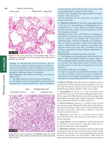
           Figure 17.7  Lobar pneumonia, acute congestion stage. There is  i) Macrophages are the predominant cells in the alveolar
           congestion of septal walls while the air spaces contain pale oedema  spaces, while neutrophils diminish in number. Many of
           fluid and a few red cells.
                                                                 the macrophages contain engulfed neutrophils and debris.
            Grossly, the affected lobe is firm and heavy. The cut  ii) Granular and fragmented strands of fibrin in the
            surface is dry, granular and grey in appearance with liver-  alveolar spaces are seen due to progressive enzymatic
            like consistency (Fig. 17.9, A). The change in colour from  digestion.
            red to grey begins at the hilum and spreads towards the  iii) Alveolar capillaries are engorged.
     SECTION III
            periphery. Fibrinous pleurisy is prominent.          iv) There is progressive removal of fluid content as well
            Histologically, the following changes are present    as cellular exudate from the air spaces, partly by
            (Fig. 17.9,B):                                       expectoration but mainly by lymphatics, resulting in
            i) The fibrin strands are dense and more numerous.   restoration of normal lung parenchyma with aeration.
            ii) The cellular exudate of neutrophils is reduced due to  COMPLICATIONS. Since the advent of antibiotics, serious
            disintegration of many inflammatory cells as evidenced
                                                               complications of lobar pneumonia are uncommon. However,
                                                               they may develop in neglected cases and in patients with
                                                               impaired immunologic defenses. These are as under:
                                                               1. Organisation. In about 3% of cases, resolution of the
                                                               exudate does not occur but instead it undergoes organisation.
                                                               There is ingrowth of fibroblasts from the alveolar septa resul-
     Systemic Pathology
                                                               ting in fibrosed, tough, airless leathery lung tissue. This type
                                                               of post-pneumonic fibrosis is called carnification.
                                                               2. Pleural effusion. About 5% of treated cases of lobar
                                                               pneumonia develop inflammation of the pleura with effusion.
                                                               The pleural effusion usually resolves but sometimes may
                                                               undergo organisation with fibrous adhesions between
                                                               visceral and parietal pleura.
                                                               3. Empyema. Less than 1% of treated cases of lobar
                                                               pneumonia develop encysted pus in the pleural cavity
                                                               termed empyema.
                                                               4. Lung abscess. A rare complication of lobar pneumonia
                                                               is formation of lung abscess, especially when there is
                                                               secondary infection by other organisms.
                                                               5. Metastatic infection. Occasionally, infection in the lungs
                                                               and pleural cavity in lobar pneumonia may extend into the
                                                               pericardium and the heart causing purulent pericarditis,
           Figure 17.8  Lobar pneumonia, red hepatisation stage. The alveoli
           are filled with cellular exudates composed of neutrophils admixed with  bacterial endocarditis and myocarditis. Other forms of
           some red cells.                                     metastatic infection encountered rarely in lobar pneumonias

