


Page 488 - Textbook of Pathology, 6th Edition
P. 488
472


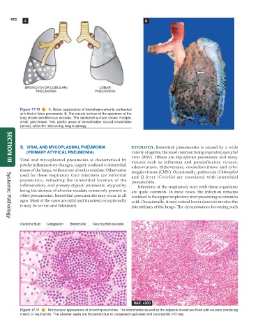
           Figure 17.10  A, Gross appearance of bronchopneumonia contrasted
           with that of lobar pneumonia. B, The pleural surface of the specimen of the
           lung shows serofibrinous exudate. The sectioned surface shows multiple,
           small, grey-brown, firm, patchy areas of consolidation around bronchioles
           (arrow). while the intervening lung is spongy.


           B. VIRAL AND MYCOPLASMAL PNEUMONIA                  ETIOLOGY. Interstitial pneumonitis is caused by a wide
              (PRIMARY ATYPICAL PNEUMONIA)                     variety of agents, the most common being respiratory syncytial
                                                               virus (RSV). Others are  Mycoplasma pneumoniae and many
           Viral and mycoplasmal pneumonia is characterised by  viruses such as influenza and parainfluenza viruses,
     SECTION III
           patchy inflammatory changes, largely confined to interstitial  adenoviruses, rhinoviruses, coxsackieviruses and cyto-
           tissue of the lungs, without any alveolar exudate. Other terms
                                                               megaloviruses (CMV). Occasionally, psittacosis (Chlamydia)
           used for these respiratory tract infections are interstitial  and Q fever  (Coxiella)  are associated with interstitial
           pneumonitis, reflecting the interstitial location of the  pneumonitis.
           inflammation, and  primary atypical pneumonia, atypicality  Infections of the respiratory tract with these organisms
           being the absence of alveolar exudate commonly present in  are quite common. In most cases, the infection remains
           other pneumonias. Interstitial pneumonitis may occur in all  confined to the upper respiratory tract presenting as common
           ages. Most of the cases are mild and transient; exceptionally  cold. Occasionally, it may extend lower down to involve the
           it may be severe and fulminant.                     interstitium of the lungs. The circumstances favouring such


     Systemic Pathology


           Figure 17.11  Microscopic appearance of bronchopneumonia. The bronchioles as well as the adjacent alveoli are filled with exudate consisting
           chiefly of neutrophils. The alveolar septa are thickened due to congested capillaries and neutrophilic infiltrate.

