


Page 489 - Textbook of Pathology, 6th Edition
P. 489
473
TABLE 17.2: Contrasting Features of Lobar Pneumonia and Bronchopneumonia.
Feature Lobar Pneumonia Bronchopneumonia
1. Definition Acute bacterial infection of a part of a lobe Acute bacterial infection of the terminal
of one or both lungs, or the entire lobe/s bronchioles extending into adjoining alveoli
2. Age group More common in adults Commoner at extremes of age–infants and old age
3. Predisposing factors More often affects healthy individuals Preexisting diseases e.g. chronic debility, terminal
illness, flu, measles
4. Common etiologic agents Pneumococci, Klebsiella pneumoniae, Staphylococci, streptococci, Pseudomonas,
staphylococci, streptococci Haemophilus influenzae
5. Pathologic features Typical case passes through stages of Patchy consolidation with central
congestion (1-2 days) , early (2-4 days) and granularity, alveolar exudation,
late consolidation (4-8 days), followed thickened septa
by resolution (1-3 weeks)
6. Investigations Neutrophilic leucocytosis, positive blood Neutrophilic leucocytosis, positive blood culture,
culture, X-ray shows consolidation X-ray shows mottled focal opacities
7. Prognosis Better response to treatment, resolution Response to treatment variable,
common, prognosis good organisation may occur, prognosis poor
8. Complications Less common; pleural effusion, empyema, Bronchiectasis may occur; other complications
lung abscess, organisation same as for lobar pneumonia
extension of infection are malnutrition, chronic debilitating COMPLICATIONS. The major complication of interstitial
diseases and alcoholism. pneumonitis is superimposed bacterial infection and its
complications. Most cases of interstitial pneumonitis recover CHAPTER 17
MORPHOLOGIC FEATURES. Irrespective of the etio- completely. In more severe cases, there may be interstitial
logic agent, the pathologic changes are similar in all cases. fibrosis and permanent damage.
Grossly, depending upon the severity of infection, the CLINICAL FEATURES. Majority of cases of interstitial
involvement may be patchy to massive and widespread pneumonitis initially have upper respiratory symptoms with
consolidation of one or both the lungs. The lungs are fever, headache and muscle-aches. A few days later appears
heavy, congested and subcrepitant. Sectioned surface of dry, hacking, non-productive cough with retrosternal
the lung exudes small amount of frothy or bloody fluid. burning due to tracheitis and bronchitis. Blood film shows
The pleural reaction is usually infrequent and mild.
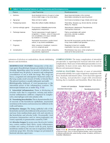
Histologically, hallmark of viral pneumonias is the
interstitial nature of the inflammatory reaction. The The Respiratory System
microscopic features are as under (Fig. 17.12):
i) Interstitial inflammation: There is thickening of
alveolar walls due to congestion, oedema and mono-
nuclear inflammatory infiltrate comprised by lympho-
cytes, macrophages and some plasma cells.
ii) Necrotising bronchiolitis: This is characterised by foci
of necrosis of the bronchiolar epithelium, inspissated
secretions in the lumina and mononuclear infiltrate in the
walls and lumina.
iii) Reactive changes: The lining epithelial cells of the
bronchioles and alveoli proliferate in the presence of virus
and may form multinucleate giant cells and syncytia in
the bronchiolar and alveolar walls. Occasionally, viral
inclusions (intranuclear and/or intracytoplasmic) are
found, especially in pneumonitis caused by CMV.
iv) Alveolar changes: In severe cases, the alveolar lumina
may contain oedema fluid, fibrin, scanty inflammatory
exudate and coating of alveolar walls by pink, hyaline
membrane similar to the one seen in respiratory distress Figure 17.12 Microscopic appearance of interstitial pneumonitis
syndrome (page 462). Alveolar changes are prominent if (viral pneumonia). There is necrotising bronchiolitis, reactive hyperplasia
bacterial infection supervenes. of alveolar epithelial cells, some having nuclear inclusions, and there is
interstitial inflammation.

