


Page 507 - Textbook of Pathology, 6th Edition
P. 507
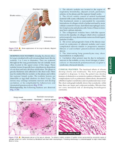
1. The silicotic nodules are located in the region of  491
                                                                 respiratory bronchioles, adjacent alveoli, pulmonary
                                                                 arteries, in the pleura and the regional lymph nodes.
                                                                 2. The  silicotic nodules consist of central hyalinised
                                                                 material with scanty cellularity and some amount of dust.
                                                                 The hyalinised centre is surrounded by concentric
                                                                 laminations of collagen which is further enclosed by more
                                                                 cellular connective tissue, dust-filled macrophages and a
                                                                 few lymphocytes and plasma cells. Some of these nodules
                                                                 may have calcium deposits.
                                                                 3. The collagenous nodules have cleft-like spaces
                                                                 between the lamellae of collagen which when examined
                                                                 polariscopically may demonstrate numerous birefringent
                                                                 particles of silica.
                                                                 4. The severe and progressive form of the disease may
                                                                 result in coalescence of adjacent nodules and cause
                                                                 complicated silicosis similar to progressive massive
           Figure 17.29  Gross appearance of the lung in silicosis, diagram-  fibrosis of coal-workers’ pneumoconiosis (described
           matic appearance.                                     above).
                                                                 5. The intervening lung parenchyma may show
            MORPHOLOGIC FEATURES. Grossly, the chronic silico-   hyperinflation or emphysema.
            tic lung is studded with well-circumscribed, hard, fibrotic  6. Cavitation when present may be due to ischaemic
            nodules, 1 to 5 mm in diameters. They are scattered  necrosis in the nodules, or may reveal changes of tuber-
            throughout the lung parenchyma but are initially more  culosis or rheumatoid pneumoconiosis (Caplan’s
            often located in the upper zones of the lungs. These  syndrome), discussed already.                       CHAPTER 17
            nodular lesions frequently have simultaneous deposition
            of coal-dust and may develop calcification. The pleura is  CLINICAL FEATURES.  The functional effects of silicosis
            grossly thickened and adherent to the chest wall. There  develop slowly and insidiously. The main presenting
            may be similar fibrotic nodules on the pleura and within  complaint is dyspnoea. In time, the patient may develop
            the regional lymph nodes. The nodular lesions are  features of obstructive or restrictive pattern of disease. Other
            detectable as egg-shell shadows in chest X-rays. The  complications such as pulmonary tuberculosis, rheumatoid
            lesions may undergo ischaemic necrosis and develop  arthritis (Caplan’s syndrome) and cor pulmonale may occur.
            cavitation, or be complicated by tuberculosis and  The chest radiograph initially shows fine nodularity, while
            rheumatoid pneumoconiosis (Fig. 17.29).            later there are larger and coalescent nodules. Silicosis does
            Histologically, the following features are observed  not carry increased risk of developing bronchogenic
            (Fig. 17.30):                                      carcinoma.                                             The Respiratory System


           Figure 17.30  Microscopic picture of the lung in silicosis. The silicotic nodule consists of hyaline centre surrounded by concentric layers of
           collagen which are further enclosed by fibroblasts and dust-laden macrophages. Polarising microscopy in photomicrograph on right shows bright
           fibres of silica.

