


Page 503 - Textbook of Pathology, 6th Edition
P. 503
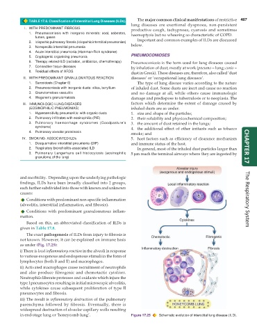
TABLE 17.8. Classification of Interstitial Lung Diseases (ILDs). The major common clinical manifestations of restrictive 487
lung diseases are exertional dyspnoea, non-persistent
I. WITH PREDOMINANT FIBROSIS productive cough, tachypnoea, cyanosis and sometimes
1. Pneumoconiosis with inorganic minerals: coal, asbestos, haemoptysis but no wheezing so characteristic of COPD.
fumes, gases
2. Idiopathic pulmonary fibrosis (Idiopathic interstitial pneumonias) Important and common examples of ILDs are discussed
3. Nonspecific interstitial pneumonia below.
4. Acute interstitial pneumonia (Hamman-Rich syndrome)
5. Cryptogenic organising pneumonia PNEUMOCONIOSES
6. Therapy related-ILD (radiation, antibiotics, chemotherapy) Pneumoconiosis is the term used for lung diseases caused
7. Connective tissue diseases by inhalation of dust, mostly at work (pneumo = lung; conis =
8. Residual effects of ARDS dust in Greek). These diseases are, therefore, also called ‘dust
II. WITH PREDOMINANT GRANULOMATOUS REACTION diseases’ or ‘occupational lung diseases’.
1. Sarcoidosis (Chapter 6) The type of lung disease varies according to the nature
2. Pneumoconiosis with inorganic dusts: silica, beryllium of inhaled dust. Some dusts are inert and cause no reaction
3. Granulomatous vasculitis and no damage at all, while others cause immunologic
4. Wegener’s granulomatosis damage and predispose to tuberculosis or to neoplasia. The
III. IMMUNOLOGIC LUNG DISEASES factors which determine the extent of damage caused by
(EOSINOPHILIC PNEUMONIAS) inhaled dusts are as under:
1. Hypersensitivity pneumonitis: with organic dusts 1. size and shape of the particles;
2. Pulmonary infiltrates with eosinophilia (PIE) 2. their solubility and physicochemical composition;
3. Pulmonary haemorrhage syndromes (Goodpasture’s 3. the amount of dust retained in the lungs;
syndrome) 4. the additional effect of other irritants such as tobacco
4. Pulmonary alveolar proteinosis
smoke; and
IV. SMOKING- ASSOCIATED ILDs 5. host factors such as efficiency of clearance mechanism
1. Desquamative interstitial pneumonia (DIP) and immune status of the host.
2. Respiratory bronchiolitis-associated ILD In general, most of the inhaled dust particles larger than CHAPTER 17
3. Pulmonary Langerhans cell histiocytosis (eosinophilic 5 μm reach the terminal airways where they are ingested by
granuloma of the lung)
and morbidity. Depending upon the underlying pathologic
findings, ILDs have been broadly classified into 2 groups,
each further subdivided into those with known and unknown
causes:
Conditions with predominant non-specific inflammation The Respiratory System
(alveolitis, interstitial inflammation, and fibrosis).
Conditions with predominant granulomatous inflam-
mation.
Based on this, an abbreviated classification of ILDs is
given in Table 17.8.
The exact pathogenesis of ILDs from injury to fibrosis is
not known. However, it can be explained on immune basis
as under (Fig. 17.25):
i) There is local inflammatory reaction in the alveoli in response
to various exogenous and endogenous stimuli in the form of
lymphocytes (both B and T) and macrophages.
ii) Activated macrophages cause recruitment of neutrophils
and also produce fibrogenic and chemotactic cytokines.
Neutrophils liberate proteases and oxidants which injure the
type I pneumocytes resulting in initial microscopic alveolitis,
while cytokines cause subsequent proliferation of type II
pneumocytes and fibrosis.
iii) The result is inflammatory destruction of the pulmonary
parenchyma followed by fibrosis. Eventually, there is
widespread destruction of alveolar capillary walls resulting
in end-stage lung or ‘honeycomb lung’. Figure 17.25 Schematic evolution of interstitial lung disease (ILD).

