


Page 502 - Textbook of Pathology, 6th Edition
P. 502
486
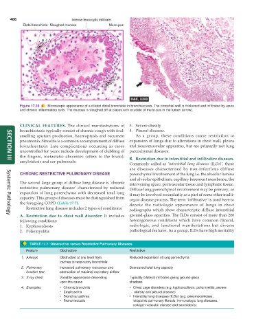
Figure 17.24 Microscopic appearance of a dilated distal bronchiole in bronchiectasis. The bronchial wall is thickened and infiltrated by acute
and chronic inflammatory cells. The mucosa is sloughed off at places with exudate of muco-pus in the lumen (arrow).
CLINICAL FEATURES. The clinical manifestations of 3. Severe obesity
bronchiectasis typically consist of chronic cough with foul- 4. Pleural diseases.
smelling sputum production, haemoptysis and recurrent As a group, these conditions cause restriction to
pneumonia. Sinusitis is a common accompaniment of diffuse expansion of lungs due to alterations in chest wall, pleura
bronchiectasis. Late complications occurring in cases and neuromuscular apparatus, but are primarily not lung
uncontrolled for years include development of clubbing of parenchymal diseases.
the fingers, metastatic abscesses (often to the brain), B. Restriction due to interstitial and infiltrative diseases.
amyloidosis and cor pulmonale.
SECTION III
Commonly called as ‘interstitial lung diseases (ILDs)’, these
are diseases characterised by non-infectious diffuse
CHRONIC RESTRICTIVE PULMONARY DISEASE parenchymal involvement of the lung i.e. the alveolar lumina
and alveolar epithelium, capillary basement membrane, the
The second large group of diffuse lung disease is ‘chronic intervening space, perivascular tissue and lymphatic tissue.
restrictive pulmonary disease’ characterised by reduced Diffuse lung parenchymal involvement may be primary, or
expansion of lung parenchyma with decreased total lung it may be involved secondarily as a part of some other multi-
capacity. This group of diseases must be distinguished from organ disease process. The term ‘infiltrative’ is used here to
the foregoing COPD (Table 17.7). denote the radiologic appearance of lungs in chest
Restrictive lung disease includes 2 types of conditions: radiographs which show characteristic diffuse interstitial
A. Restriction due to chest wall disorder: It includes ground-glass opacities. The ILDs consist of more than 200
Systemic Pathology
following conditions: heterogeneous conditions which have common clinical,
1. Kyphoscoliosis radiologic, and functional manifestations but diverse
2. Poliomyelitis pathological features. As a group, ILDs have high mortality
TABLE 17.7: Obstructive versus Restrictive Pulmonary Diseases.
Feature Obstructive Restrictive
1. Airways Obstructed at any level from Reduced expansion of lung parenchyma
trachea to respiratory bronchiole
2. Pulmonary Increased pulmonary resistance and Decreased total lung capacity
function test obstruction of maximal expiratory airflow
3. X-ray chest Variable appearance depending Typically bilateral infiltrates giving ground-glass
upon the cause shadows
4. Examples • Chronic bronchitis • Chest cage disorders (e.g. kyphoscoliosis, poliomyelitis, severe
• Emphysema obesity and pleural disease)
• Bronchial asthma • Interstitial lung diseases (ILDs) (e.g. pneumoconioses,
• Bronchiectasis idiopathic pulmonary fibrosis, immunologic lung diseases,
collagen-vascular disease and sarcoidosis)

