


Page 505 - Textbook of Pathology, 6th Edition
P. 505
489


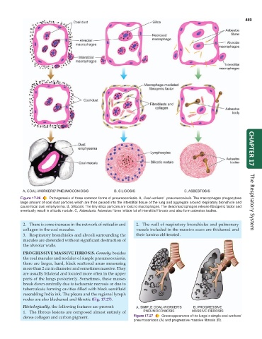
           Figure 17.26  Pathogenesis of three common forms of pneumoconiosis. A, Coal-workers’  pneumoconiosis. The macrophages phagocytose  CHAPTER 17
           large amount of coal dust particles which are then passed into the interstitial tissue of the lung and aggregate around respiratory bronchiole and  The Respiratory System
           cause focal dust emphysema. B, Silicosis. The tiny silica particles are toxic to macrophages. The dead macrophages release fibrogenic factor and
           eventually result in silicotic nodule. C, Asbestosis. Asbestos fibres initiate lot of interstitial fibrosis and also form asbestos bodies.


            2. There is some increase in the network of reticulin and  2. The wall of respiratory bronchioles and pulmonary
            collagen in the coal macules.                        vessels included in the massive scars are thickened and
            3. Respiratory bronchioles and alveoli surrounding the  their lumina obliterated.
            macules are distended without significant destruction of
            the alveolar walls.
            PROGRESSIVE MASSIVE FIBROSIS. Grossly, besides
            the coal macules and nodules of simple pneumoconiosis,
            there are larger, hard, black scattered areas measuring
            more than 2 cm in diameter and sometimes massive. They
            are usually bilateral and located more often in the upper
            parts of the lungs posteriorly. Sometimes, these masses
            break down centrally due to ischaemic necrosis or due to
            tuberculosis forming cavities filled with black semifluid
            resembling India ink. The pleura and the regional lymph
            nodes are also blackened and fibrotic (Fig. 17.27).
            Histologically, the following features are present:
            1. The fibrous lesions are composed almost entirely of
            dense collagen and carbon pigment.                 Figure 17.27  Gross appearance of the lungs in simple coal-workers’
                                                               pneumoconiosis (A) and progressive massive fibrosis (B).

