


Page 504 - Textbook of Pathology, 6th Edition
P. 504
488 asymptomatic accumulation of carbon dust in the lungs of
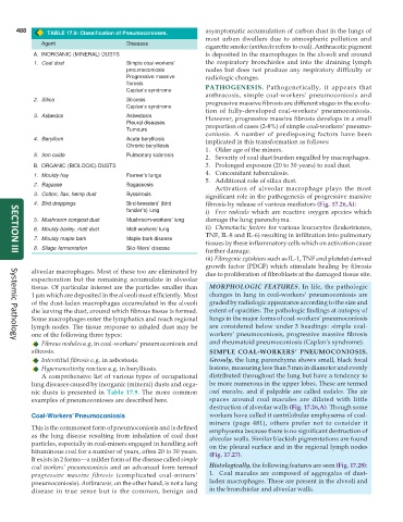
TABLE 17.9: Classification of Pneumoconioses.
most urban dwellers due to atmospheric pollution and
Agent Diseases
cigarette smoke (anthacite refers to coal). Anthracotic pigment
A. INORGANIC (MINERAL) DUSTS is deposited in the macrophages in the alveoli and around
1. Coal dust Simple coal-workers’ the respiratory bronchioles and into the draining lymph
pneumoconiosis nodes but does not produce any respiratory difficulty or
Progressive massive radiologic changes.
fibrosis PATHOGENESIS. Pathogenetically, it appears that
Caplan’s syndrome
anthracosis, simple coal-workers’ pneumoconiosis and
2. Silica Silicosis progressive massive fibrosis are different stages in the evolu-
Caplan’s syndrome
tion of fully-developed coal-workers’ pneumoconiosis.
3. Asbestos Asbestosis However, progressive massive fibrosis develops in a small
Pleural diseases proportion of cases (2-8%) of simple coal-workers’ pneumo-
Tumours
coniosis. A number of predisposing factors have been
4. Beryllium Acute berylliosis implicated in this transformation as follows:
Chronic berylliosis
1. Older age of the miners.
5. Iron oxide Pulmonary siderosis 2. Severity of coal dust burden engulfed by macrophages.
B. ORGANIC (BIOLOGIC) DUSTS 3. Prolonged exposure (20 to 30 years) to coal dust.
4. Concomitant tuberculosis.
1. Mouldy hay Farmer’s lungs
5. Additional role of silica dust.
2. Bagasse Bagassosis
Activation of alveolar macrophage plays the most
3. Cotton, flax, hemp dust Byssinosis significant role in the pathogenesis of progressive massive
4. Bird droppings Bird-breeders’ (bird fibrosis by release of various mediators (Fig. 17.26,A):
fancier’s) lung i) Free radicals which are reactive oxygen species which
5. Mushroom compost dust Mushroom-workers’ lung damage the lung parenchyma.
6. Mouldy barley, malt dust Malt-workers’ lung ii) Chemotactic factors for various leucocytes (leukotrienes,
TNF, IL-8 and IL-6) resulting in infiltration into pulmonary
7. Mouldy maple bark Maple-bark disease
tissues by these inflammatory cells which on activation cause
8. Silage fermentation Silo-fillers’ disease
further damage.
SECTION III
iii) Fibrogenic cytokines such as IL-1, TNF and platelet derived
growth factor (PDGF) which stimulate healing by fibrosis
alveolar macrophages. Most of these too are eliminated by due to proliferation of fibroblasts at the damaged tissue site.
expectoration but the remaining accumulate in alveolar
tissue. Of particular interest are the particles smaller than MORPHOLOGIC FEATURES. In life, the pathologic
1 μm which are deposited in the alveoli most efficiently. Most changes in lung in coal-workers’ pneumoconiosis are
of the dust-laden macrophages accumulated in the alveoli graded by radiologic appearance according to the size and
die leaving the dust, around which fibrous tissue is formed. extent of opacities. The pathologic findings at autopsy of
Some macrophages enter the lymphatics and reach regional lungs in the major forms of coal-workers’ pneumoconiosis
lymph nodes. The tissue response to inhaled dust may be are considered below under 3 headings: simple coal-
one of the following three types: workers’ pneumoconiosis, progressive massive fibrosis
Fibrous nodules e.g. in coal-workers’ pneumoconiosis and and rheumatoid pneumoconiosis (Caplan’s syndrome).
Systemic Pathology
silicosis. SIMPLE COAL-WORKERS’ PNEUMOCONIOSIS.
Interstitial fibrosis e.g. in asbestosis. Grossly, the lung parenchyma shows small, black focal
Hypersensitivity reaction e.g. in berylliosis. lesions, measuring less than 5 mm in diameter and evenly
A comprehensive list of various types of occupational distributed throughout the lung but have a tendency to
lung diseases caused by inorganic (mineral) dusts and orga- be more numerous in the upper lobes. These are termed
nic dusts is presented in Table 17.9. The more common coal macules, and if palpable are called nodules. The air
examples of pneumoconioses are described here. spaces around coal macules are dilated with little
destruction of alveolar walls (Fig. 17.26,A). Though some
Coal-Workers’ Pneumoconiosis workers have called it centrilobular emphysema of coal-
miners (page 481), others prefer not to consider it
This is the commonest form of pneumoconiosis and is defined emphysema because there is no significant destruction of
as the lung disease resulting from inhalation of coal dust alveolar walls. Similar blackish pigmentations are found
particles, especially in coal-miners engaged in handling soft on the pleural surface and in the regional lymph nodes
bituminous coal for a number of years, often 20 to 30 years. (Fig. 17.27).
It exists in 2 forms—a milder form of the disease called simple
coal workers’ pneumoconiosis and an advanced form termed Histologically, the following features are seen (Fig. 17.28):
progressive massive fibrosis (complicated coal-miners’ 1. Coal macules are composed of aggregates of dust-
pneumoconiosis). Anthracosis, on the other hand, is not a lung laden macrophages. These are present in the alveoli and
disease in true sense but is the common, benign and in the bronchiolar and alveolar walls.

