
Page 660 - Textbook of Pathology, 6th Edition
P. 660
644 Histologically, the following patterns are observed:
1. Most gallbladder cancers are adenocarcinomas (90%).
They may be papillary or infiltrative, well-differentiated
or poorly-differentiated. Most are non-mucin secreting but
some are colloid carcinomas forming mucus pools.
2. About 5% of gallbladder cancers are squamous cell
carcinomas arising from squamous metaplastic epithelium.
3. A few cases show both squamous and adeno-
carcinoma pattern of growth called adenosquamous
carcinoma.
CLINICAL FEATURES. Carcinoma of the gallbadder is
slow-growing and causes symptoms late in the course of
disease. Quite often, the diagnosis is made when gallbladder
is removed for cholelithiasis. The symptomatic cases have
pain, jaundice, noticeable mass, anorexia and weight loss.
In such case, the growth has usually invaded the liver and
other adjacent organs and has metastasised to regional lymph
nodes and more distant sites such as the lung, peritoneum
and gastrointestinal tract.
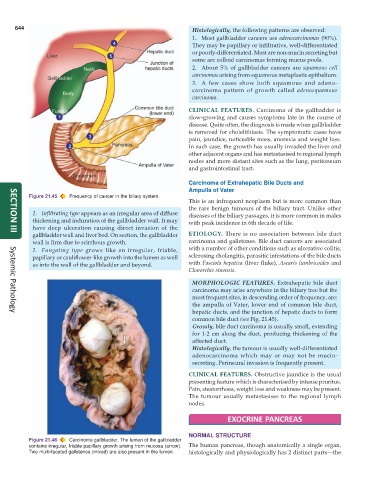
Carcinoma of Extrahepatic Bile Ducts and
Ampulla of Vater
Figure 21.45 Frequency of cancer in the biliary system.
This is an infrequent neoplasm but is more common than
the rare benign tumours of the biliary tract. Unlike other
1. Infiltrating type appears as an irregular area of diffuse diseases of the biliary passages, it is more common in males
thickening and induration of the gallbladder wall. It may with peak incidence in 6th decade of life.
have deep ulceration causing direct invasion of the
gallbladder wall and liver bed. On section, the gallbladder ETIOLOGY. There is no association between bile duct
SECTION III
wall is firm due to scirrhous growth. carcinoma and gallstones. Bile duct cancers are associated
2. Fungating type grows like an irregular, friable, with a number of other conditions such as ulcerative colitis,
papillary or cauliflower-like growth into the lumen as well sclerosing cholangitis, parasitic infestations of the bile ducts
as into the wall of the gallbladder and beyond. with Fasciola hepatica (liver fluke), Ascaris lumbricoides and
Clonorchis sinensis.
MORPHOLOGIC FEATURES. Extrahepatic bile duct
carcinoma may arise anywhere in the biliary tree but the
most frequent sites, in descending order of frequency, are:
the ampulla of Vater, lower end of common bile duct,
hepatic ducts, and the junction of hepatic ducts to form
Systemic Pathology
common bile duct (see Fig. 21.45).
Grossly, bile duct carcinoma is usually small, extending
for 1-2 cm along the duct, producing thickening of the
affected duct.
Histologically, the tumour is usually well-differentiated
adenocarcinoma which may or may not be mucin-
secreting. Perineural invasion is frequently present.
CLINICAL FEATURES. Obstructive jaundice is the usual
presenting feature which is characterised by intense pruritus.
Pain, steatorrhoea, weight loss and weakness may be present.
The tumour usually metastasises to the regional lymph
nodes.
EXOCRINE PANCREAS
NORMAL STRUCTURE
Figure 21.46 Carcinoma gallbladder. The lumen of the gallbladder
contains irregular, friable papillary growth arising from mucosa (arrow). The human pancreas, though anatomically a single organ,
Two multi-faceted gallstones (mixed) are also present in the lumen. histologically and physiologically has 2 distinct parts—the