
Page 672 - Textbook of Pathology, 6th Edition
P. 672
656 to back pressure. Radiologically, uraemic pneumonitis shows
characteristic central, butterfly-pattern of oedema and
congestion in the chest radiograph.
5. Digestive system. Azotaemia directly induces mucosal
ulcerations in the lining of the stomach and intestines.
Subsequent bleeding can aggravate the existing anaemia.
Gastrointestinal irritation may cause nausea, vomiting and
diarrhoea.
6. Skeletal system. The skeletal manifestations of renal
failure are referred to as renal osteodystrophy (Chapter 28).
Two major types of skeletal disorders may occur:
i) Osteomalacia occurs from deficiency of a form of vitamin
D which is normally activated by the kidney (page 248). Since
vitamin D is essential for absorption of calcium, its deficiency
results in inadequate deposits of calcium in bone tissue.
ii) Osteitis fibrosa occurs due to elevated levels of
parathormone. How parathormone excess develops in CRF
is complex. As the GFR is decreased, increasing levels of
phosphates accumulate in the extracellular fluid which, in
turn, cause decline in calcium levels. Decreased calcium level
triggers the secretion of parathormone which mobilises
calcium from bone and increases renal tubular reabsorption
of calcium thereby conserving it. However, if the process of
resorption of calcium phosphate from bone continues for
sufficient time, hypercalcaemia may be induced with
deposits of excess calcium salts in joints and soft tissues and
weakening of bones (renal osteodystrophy).
SECTION III
CONGENITAL MALFORMATIONS
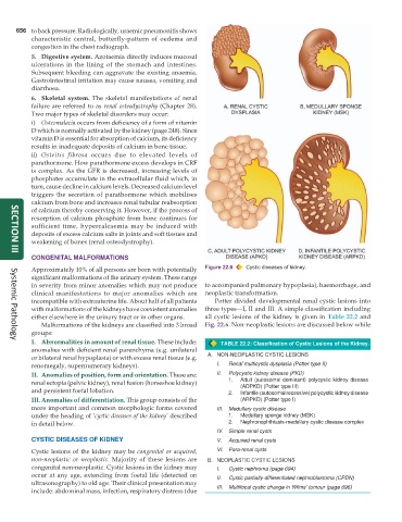
Approximately 10% of all persons are born with potentially Figure 22.6 Cystic diseases of kidney.
significant malformations of the urinary system. These range
in severity from minor anomalies which may not produce to accompanied pulmonary hypoplasia), haemorrhage, and
clinical manifestations to major anomalies which are neoplastic transformation.
incompatible with extrauterine life. About half of all patients Potter divided developmental renal cystic lesions into
with malformations of the kidneys have coexistent anomalies three types—I, II and III. A simple classification including
either elsewhere in the urinary tract or in other organs. all cystic lesions of the kidney is given in Table 22.2 and
Malformations of the kidneys are classified into 3 broad Fig. 22.6. Non-neoplastic lesions are discussed below while
groups:
I. Abnormalities in amount of renal tissue. These include: TABLE 22.2: Classification of Cystic Lesions of the Kidney.
Systemic Pathology
anomalies with deficient renal parenchyma (e.g. unilateral
or bilateral renal hypoplasia) or with excess renal tissue (e.g. A. NON-NEOPLASTIC CYSTIC LESIONS
renomegaly, supernumerary kidneys). I. Renal multicystic dysplasia (Potter type II)
II. Anomalies of position, form and orientation. These are: II. Polycystic kidney disease (PKD)
renal ectopia (pelvic kidney), renal fusion (horseshoe kidney) 1. Adult (autosomal dominant) polycystic kidney disease
(ADPKD) (Potter type III)
and persistent foetal lobation. 2. Infantile (autosomal recessive) polycystic kidney disease
III. Anomalies of differentiation. This group consists of the (ARPKD) (Potter type I)
more important and common morphologic forms covered III. Medullary cystic disease
under the heading of ‘cystic diseases of the kidney’ described 1. Medullary sponge kidney (MSK)
in detail below. 2. Nephronophthiasis-medullary cystic disease complex
IV. Simple renal cysts
CYSTIC DISEASES OF KIDNEY V. Acquired renal cysts
Cystic lesions of the kidney may be congenital or acquired, VI. Para-renal cysts
non-neoplastic or neoplastic. Majority of these lesions are B. NEOPLASTIC CYSTIC LESIONS
congenital non-neoplastic. Cystic lesions in the kidney may I. Cystic nephroma (page 694)
occur at any age, extending from foetal life (detected on II. Cystic partially-differentiated nephroblastoma (CPDN)
ultrasonography) to old age. Their clinical presentation may
include: abdominal mass, infection, respiratory distress (due III. Multifocal cystic change in Wilms’ tumour (page 696)