
Page 718 - Textbook of Pathology, 6th Edition
P. 718
702
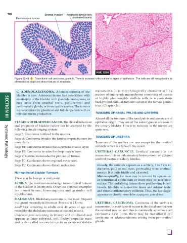
Figure 22.48 Transitional cell carcinoma, grade II. There is increase in the number of layers of epithelium. The cells are still recognisable as
of transitional origin and show features of anaplasia.
C. ADENOCARCINOMA. Adenocarcinoma of the myosarcoma. It is morphologically characterised by
bladder is rare. Adenocarcinoma has association with masses of embryonic mesenchyme consisting of masses
exostrophy of the bladder with glandular metaplasia, or of highly pleomorphic stellate cells in myxomatous
may arise from urachal rests, periurethral and background. Similar tumours occur in the female genital
periprostatic glands, or from cystitis cystica. The tumour tract (Chapter 24).
is characterised by glandular and tubular pattern with or
without mucus production. TUMOURS OF RENAL PELVIS AND URETERS
Almost all the tumours of the renal pelvis and ureters are of
SECTION III
STAGING OF BLADDER CANCER. The clinical behaviour epithelial origin. They are of the same types as are seen in
and prognosis of bladder cancer can be assessed by the the urinary bladder. However, tumours in the ureters are
following simple staging system: quite rare.
Stage 0: Carcinoma confined to the mucosa.
TUMOURS OF URETHRA
Stage A: Carcinoma invades the lamina propria but not the
muscularis. Tumours of the urethra are rare except for the urethral
Stage B1: Carcinoma invades the superficial muscle layer. caruncle which is a tumour-like lesion.
Stage B2: Carcinoma invades the deep muscle layer. URETHRAL CARUNCLE. Urethral caruncle is not
Stage C: Carcinoma invades the perivesical tissues. uncommon. It is an inflammatory lesion present on external
urethral meatus in elderly females.
Systemic Pathology
Stage D1: Carcinoma shows regional metastases.
Stage D2: Carcinoma shows distant metastases. Grossly, the caruncle appears as a solitary, 1 to 2 cm in
diameter, pink or red mass, protruding from urethral
Non-epithelial Bladder Tumours meatus. It is quite friable and ulcerated.
Microscopically, the mass may be covered by squamous
These may be benign or malignant.
or transitional epithelium or there may be ulcerated
BENIGN. The most common benign mesenchymal tumour surface. The underlying tissues show proliferating blood
of the bladder is leiomyoma. Other less common examples vessels, fibroblastic connective tissue and intense acute
are neurofibroma, haemangioma and granular cell and chronic inflammatory infiltrate. Thus, the histologic
myoblastoma. appearance closely resembles a pyogenic granuloma.
MALIGNANT. Rhabdomyosarcoma is the most frequent
malignant mesenchymal tumour. It exists in 2 forms: URETHRAL CARCINOMA. Carcinoma of the urethra is
Adult form occurring in adults over 40 years of age and uncommon. In most cases it occurs in the distal urethra near
resembles the rhabdomyosarcoma of skeletal muscle. the external meatus and thus is commonly squamous cell
Childhood form occurring in infancy and childhood and carcinoma. Less often, there may be transitional cell
appears as large polypoid, soft, fleshy, grapelike mass carcinoma or adenocarcinoma arising from periurethral
and is also called sarcoma botryoides or embryonal rhabdo- glands.
❑