
Page 722 - Textbook of Pathology, 6th Edition
P. 722
706 Histologically, it consists of a granuloma composed of
histiocytes, epithelioid cells, lymphocytes and some
neutrophils. Characteristically, the centre of spermatic
granuloma contains spermatozoa and necrotic debris. The
late lesions have fibroblastic proliferation at the periphery
and hyalinisation.
Elephantiasis
Elephantiasis is enormous thickening of the scrotal skin
resembling the elephant’s hide and results in enlargement
of the scrotum. The condition results from filariasis in which
the adult worm lives in the lymphatics, while the larvae
travel in the blood. The most important variety of filaria is
Wuchereria bancrofti. The condition is common in all tropical
countries. The vector is generally the Culex mosquito. The
patients may remain asymptomatic or may manifest with
fever, local pain, swelling, rash, tender lymphadenopathy
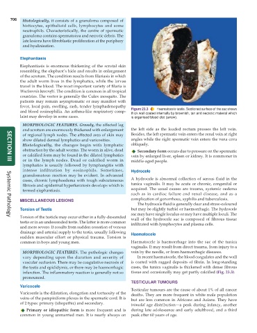
and blood eosinophilia. An asthma-like respiratory comp- Figure 23.3 Haematocele testis. Sectioned surface of the sac shows
thick wall coated internally by brownish, tan and necrotic material which
laint may develop in some cases. is organised blood clot (arrow).
MORPHOLOGIC FEATURES. Grossly, the affected leg
and scrotum are enormously thickened with enlargement the left side as the loaded rectum presses the left vein.
of regional lymph nodes. The affected area of skin may Besides, the left spermatic vein enters the renal vein at right
show dilated dermal lymphatics and varicosities. angles while the right spermatic vein enters the vena cava
Histologically, the changes begin with lymphatic obliquely.
obstruction by the adult worms. The worm in alive, dead Secondary form occurs due to pressure on the spermatic
or calcified form may be found in the dilated lymphatics vein by enlarged liver, spleen or kidney. It is commoner in
or in the lymph nodes. Dead or calcified worm in middle-aged people.
SECTION III
lymphatics is usually followed by lymphangitis with
intense infiltration by eosinophils. Sometimes, Hydrocele
granulomatous reaction may be evident. In advanced
cases, chronic lymphoedema with tough subcutaneous A hydrocele is abnormal collection of serous fluid in the
fibrosis and epidermal hyperkeratosis develops which is tunica vaginalis. It may be acute or chronic, congenital or
termed elephantiasis. acquired. The usual causes are trauma, systemic oedema
such as in cardiac failure and renal disease, and as a
MISCELLANEOUS LESIONS complication of gonorrhoea, syphilis and tuberculosis.
The hydrocele fluid is generally clear and straw-coloured
Torsion of Testis but may be slightly turbid or haemorrhagic. The hydrocele
sac may have single loculus or may have multiple loculi. The
Torsion of the testicle may occur either in a fully-descended wall of the hydrocele sac is composed of fibrous tissue
Systemic Pathology
testis or in an undescended testis. The latter is more common infiltrated with lymphocytes and plasma cells.
and more severe. It results from sudden cessation of venous
drainage and arterial supply to the testis, usually following Haematocele
sudden muscular effort or physical trauma. Torsion is
common in boys and young men. Haematocele is haemorrhage into the sac of the tunica
vaginalis. It may result from direct trauma, from injury to a
MORPHOLOGIC FEATURES. The pathologic changes vein by the needle, or from haemorrhagic diseases.
vary depending upon the duration and severity of In recent haematocele, the blood coagulates and the wall
vascular occlusion. There may be coagulative necrosis of is coated with ragged deposits of fibrin. In long-standing
the testis and epididymis, or there may be haemorrhagic cases, the tunica vaginalis is thickened with dense fibrous
infarction. The inflammatory reaction is generally not so tissue and occasionally may get partly calcified (Fig. 23.3).
pronounced.
TESTICULAR TUMOURS
Varicocele
Testicular tumours are the cause of about 1% of all cancer
Varicocele is the dilatation, elongation and tortuosity of the deaths. They are more frequent in white male population
veins of the pampiniform plexus in the spermatic cord. It is but are less common in Africans and Asians. They have
of 2 types: primary (idiopathic) and secondary. trimodal age distribution—a peak during infancy, another
Primary or idiopathic form is more frequent and is during late adolescence and early adulthood, and a third
common in young unmarried men. It is nearly always on peak after 60 years of age.