
Page 713 - Textbook of Pathology, 6th Edition
P. 713
697
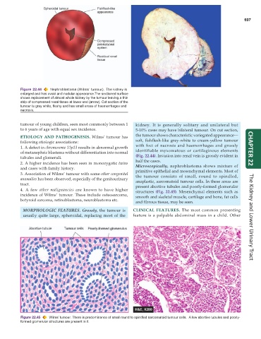
Figure 22.44 Nephroblastoma (W ilms’ tumour). The kidney is
enlarged and has ovoid and nodular appearance. The sectioned surface
shows replacement of almost whole kidney by the tumour leaving a thin
strip of compressed renal tissue at lower end (arrow). Cut section of the
tumour is gray white, fleshy and has small areas of haemorrhages and
necrosis.
tumour of young children, seen most commonly between 1 kidney. It is generally solitary and unilateral but
to 6 years of age with equal sex incidence. 5-10% cases may have bilateral tumour. On cut section,
ETIOLOGY AND PATHOGENESIS. Wilms’ tumour has the tumour shows characteristic variegated appearance—
following etiologic associations: soft, fishflesh-like grey-white to cream-yellow tumour
1. A defect in chromosome 11p13 results in abnormal growth with foci of necrosis and haemorrhages and grossly CHAPTER 22
of metanephric blastema without differentiation into normal identifiable myxomatous or cartilaginous elements
tubules and glomeruli. (Fig. 22.44). Invasion into renal vein is grossly evident in
half the cases.
2. A higher incidence has been seen in monozygotic twins Microscopically, nephroblastoma shows mixture of
and cases with family history.
primitive epithelial and mesenchymal elements. Most of
3. Association of Wilms’ tumour with some other congenital the tumour consists of small, round to spindled,
anomalies has been observed, especially of the genitourinary anaplastic, sarcomatoid tumour cells. In these areas are
tract. present abortive tubules and poorly-formed glomerular
4. A few other malignancies are known to have higher structures (Fig. 22.45). Mesenchymal elements such as
incidence of Wilms’ tumour. These include osteosarcoma, smooth and skeletal muscle, cartilage and bone, fat cells
botyroid sarcoma, retinoblastoma, neuroblastoma etc.
and fibrous tissue, may be seen.
MORPHOLOGIC FEATURES. Grossly, the tumour is CLINICAL FEATURES. The most common presenting
usually quite large, spheroidal, replacing most of the feature is a palpable abdominal mass in a child. Other The Kidney and Lower Urinary Tract
Figure 22.45 Wilms’ tumour. There is predominance of small round to spindled sarcomatoid tumour cells. A few abortive tubules and poorly-
formed glomerular structures are present in it.