
Page 721 - Textbook of Pathology, 6th Edition
P. 721
2. Acquired block e.g. due to gonorrhoea and surgical Tuberculous Epididymo-orchitis 705
intervention. Tuberculosis invariably begins in the epididymis and
3. Impaired sperm motility in the presence of normal sperm spreads to involve the testis. Tuberculous epididymo-orchitis
counts e.g. immotile cilia syndrome (Chapter 17). is generally secondary tuberculosis from elsewhere in the
body. It may occur either by direct spread from genitourinary
INFLAMMATIONS tuberculosis such as tuberculous seminal vesiculitis,
Inflammation of the testis is termed as orchitis and of prostatitis and renal tuberculosis, or may reach by
epididymis is called as epididymitis; the latter being more haematogenous spread of infection such as from tuberculosis
common. A combination epididymo-orchitis may also occur. of the lungs. Primary genital tuberculosis may occur rarely.
A few important types are described below.
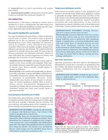
MORPHOLOGIC FEATURES. Grossly, discrete,
Non-specific Epididymitis and Orchitis yellowish, caseous necrotic areas are seen.
Microscopically, numerous tubercles which may coalesce
Non-specific epididymitis and orchitis, or their combination, to form large caseous mass are seen. Characteristics of
may be acute or chronic. The common routes of spread of typical tubercles such as epithelioid cells, peripheral
infection are via the vas deferens, or via lymphatic and mantle of lymphocytes, occasional multinucleate giant
haematogenous routes. Most frequently, the infection is cells and central areas of caseation necrosis are seen
caused by urethritis, cystitis, prostatitis and seminal (Fig. 23.2). Numerous acid-fast bacilli can be
vesiculitis. Other causes are mumps, smallpox, dengue fever, demonstrated by Ziehl-Neelsen staining. The lesions
influenza, pneumonia and filariasis. The common infecting produce extensive destruction of the epididymis and may
organisms in sexually-active men under 35 years of age are form chronic discharging sinuses on the scrotal skin. In
Neisseria gonorrhoeae and Chlamydia trachomatis, whereas in late stage, the lesions heal by fibrous scarring and may
older individuals the common organisms are urinary tract undergo calcification.
pathogens like Escherichia coli and Pseudomonas.
Spermatic Granuloma
MORPHOLOGIC FEATURES. Grossly, in acute stage the CHAPTER 23
testicle is firm, tense, swollen and congested. There may Spermatic granuloma is the term used for development of
be multiple abscesses, especially in gonorrhoeal infection. inflammatory lesions due to invasion of spermatozoa into
In chronic cases, there is usually variable degree of the stroma. Spermatic granuloma may develop due to
atrophy and fibrosis. trauma, inflammation and loss of ligature following
Histologically, acute orchitis and epididymitis are charac- vasectomy.
terised by congestion, oedema and diffuse infiltration by
neutrophils, lymphocytes, plasma cells and macrophages MORPHOLOGIC FEATURES. Grossly, the sperm granu-
or formation of neutrophilic abscesses. Acute loma is a small nodule, 3 mm to 3 cm in diameter, firm,
inflammation may resolve, or may progress to chronic white to yellowish-brown.
form. In chronic epididymo-orchitis, there is focal or
diffuse chronic inflammation, disappearance of
seminiferous tubules, fibrous scarring and destruction of
interstitial Leydig cells. Such cases usually result in
permanent sterility.
Granulomatous (Autoimmune) Orchitis The Male Reproductive System and Prostate
Non-tuberculous granulomatous orchitis is a peculiar type
of unilateral, painless testicular enlargement in middle-aged
men that may resemble a testicular tumour clinically. The
exact etiology and pathogenesis of the condition are not
known though an autoimmune basis is suspected.
MORPHOLOGIC FEATURES. Grossly, the affected testis
is enlarged with thickened tunica. Cut section of the
testicle is greyish-white to tan-brown.
Histologically, there are circumscribed non-caseating
granulomas lying within the seminiferous tubules. These
granulomas are composed of epithelioid cells, lympho-
cytes, plasma cells, some neutrophils and multinucleate
giant cells. The origin of the epithelioid cells is from Sertoli
cells lining the tubules. The tubules show peritubular Figure 23.2 Tuberculous epididymo-orchitis. The interstitium
fibrosis which merges into the interstitial tissue that is contains several epithelioid cell granulomas with central areas of
infiltrated by lymphocytes and plasma cells. caseation necrosis. These granulomas are surrounded by Langhans’
giant cells and mantle of lymphocytes.