
Page 734 - Textbook of Pathology, 6th Edition
P. 734
718
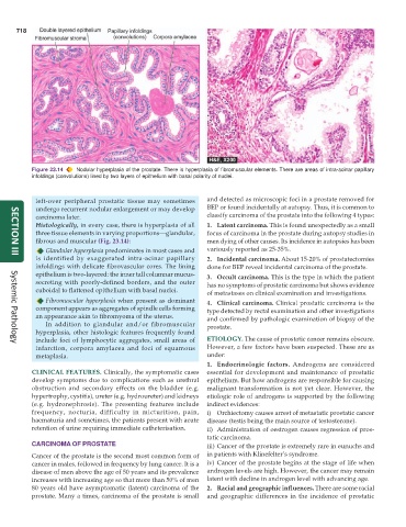
Figure 23.14 Nodular hyperplasia of the prostate. There is hyperplasia of fibromuscular elements. There are areas of intra-acinar papillary
infoldings (convolutions) lined by two layers of epithelium with basal polarity of nuclei.
left-over peripheral prostatic tissue may sometimes and detected as microscopic foci in a prostate removed for
undergo recurrent nodular enlargement or may develop BEP or found incidentally at autopsy. Thus, it is common to
carcinoma later. classify carcinoma of the prostate into the following 4 types:
Histologically, in every case, there is hyperplasia of all 1. Latent carcinoma. This is found unexpectedly as a small
three tissue elements in varying proportions—glandular, focus of carcinoma in the prostate during autopsy studies in
fibrous and muscular (Fig. 23.14): men dying of other causes. Its incidence in autopsies has been
Glandular hyperplasia predominates in most cases and variously reported as 25-35%.
is identified by exaggerated intra-acinar papillary 2. Incidental carcinoma. About 15-20% of prostatectomies
SECTION III
infoldings with delicate fibrovascular cores. The lining done for BEP reveal incidental carcinoma of the prostate.
epithelium is two-layered: the inner tall columnar mucus- 3. Occult carcinoma. This is the type in which the patient
secreting with poorly-defined borders, and the outer has no symptoms of prostatic carcinoma but shows evidence
cuboidal to flattened epithelium with basal nuclei. of metastases on clinical examination and investigations.
Fibromuscular hyperplasia when present as dominant 4. Clinical carcinoma. Clinical prostatic carcinoma is the
component appears as aggregates of spindle cells forming type detected by rectal examination and other investigations
an appearance akin to fibromyoma of the uterus. and confirmed by pathologic examination of biopsy of the
In addition to glandular and/or fibromuscular prostate.
hyperplasia, other histologic features frequently found
include foci of lymphocytic aggregates, small areas of ETIOLOGY. The cause of prostatic cancer remains obscure.
Systemic Pathology
infarction, corpora amylacea and foci of squamous However, a few factors have been suspected. These are as
metaplasia. under:
1. Endocrinologic factors. Androgens are considered
CLINICAL FEATURES. Clinically, the symptomatic cases essential for development and maintenance of prostatic
develop symptoms due to complications such as urethral epithelium. But how androgens are responsible for causing
obstruction and secondary effects on the bladder (e.g. malignant transformation is not yet clear. However, the
hypertrophy, cystitis), ureter (e.g. hydroureter) and kidneys etiologic role of androgens is supported by the following
(e.g. hydronephrosis). The presenting features include indirect evidences:
frequency, nocturia, difficulty in micturition, pain, i) Orchiectomy causes arrest of metastatic prostatic cancer
haematuria and sometimes, the patients present with acute disease (testis being the main source of testosterone).
retention of urine requiring immediate catheterisation. ii) Administration of oestrogen causes regression of pros-
tatic carcinoma.
CARCINOMA OF PROSTATE iii) Cancer of the prostate is extremely rare in eunuchs and
Cancer of the prostate is the second most common form of in patients with Klinefelter’s syndrome.
cancer in males, followed in frequency by lung cancer. It is a iv) Cancer of the prostate begins at the stage of life when
disease of men above the age of 50 years and its prevalence androgen levels are high. However, the cancer may remain
increases with increasing age so that more than 50% of men latent with decline in androgen level with advancing age.
80 years old have asymptomatic (latent) carcinoma of the 2. Racial and geographic influences. There are some racial
prostate. Many a times, carcinoma of the prostate is small and geographic differences in the incidence of prostatic