
Page 738 - Textbook of Pathology, 6th Edition
P. 738
722 Histologically, they are covered by an orderly stratified
squamous epithelium. The stroma consists of loose fibrous
and myxomatous connective tissue with some adipose
tissue and blood vessels.
Papillary Hidradenoma (Hidradenoma Papilliferum)
This is a benign tumour arising from apocrine sweat glands
of the vulva. Most commonly, it is located in the labia or in
the perianal region as a small sharply circumscribed nodule.
Histologically, the tumour lies in the dermis under a
normal epidermis. The tumour consists of papillary
structures composed of fibrovascular stalk and is covered
by double layer of epithelial cells—a layer of flattened
myoepithelial cells and an overlying layer of columnar
cells.
Condyloma Acuminatum
Condyloma acuminata or anogenital warts are benign
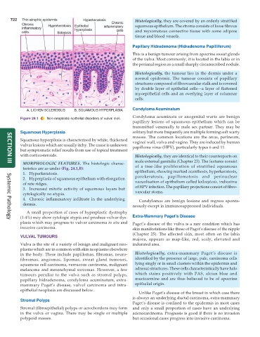
Figure 24.1 Non-neoplastic epithelial disorders of vulval skin.
papillary lesions of squamous epithelium which can be
transmitted venereally to male sex partner. They may be
Squamous Hyperplasia solitary but more frequently are multiple forming soft warty
masses. The common locations are the anus, perineum,
Squamous hyperplasia is characterised by white, thickened vaginal wall, vulva and vagina. They are induced by human
vulvar lesions which are usually itchy. The cause is unknown papilloma virus (HPV), particularly types 6 and 11.
but symptomatic relief results from use of topical treatment
with corticosteroids. Histologically, they are identical to their counterparts on
male external genitalia (Chapter 23). The features consist
MORPHOLOGIC FEATURES. The histologic charac-
SECTION III
teristics are as under (Fig. 24.1,B): of a tree-like proliferation of stratified squamous
1. Hyperkeratosis. epithelium, showing marked acanthosis, hyperkeratosis,
2. Hyperplasia of squamous epithelium with elongation parakeratosis, papillomatosis and perinuclear
of rete ridges. vacuolisation of epithelium called koilocytosis, indicative
3. Increased mitotic activity of squamous layers but of HPV infection. The papillary projections consist of fibro-
cytologically no atypia. vascular stoma.
4. Chronic inflammatory infiltrate in the underlying Condylomas are benign lesions and regress sponta-
dermis. neously except in immunosuppressed individuals.
A small proportion of cases of hyperplastic dystrophy
(1-4%) may show cytologic atypia and produce vulvar dys- Extra-Mammary Paget’s Disease
Systemic Pathology
plasia which may progress to vulvar carcinoma in situ and Paget’s disease of the vulva is a rare condition which has
invasive carcinoma. skin manifestations like those of Paget’s disease of the nipple
(Chapter 25). The affected skin, most often on the labia
VULVAL TUMOURS
majora, appears as map-like, red, scaly, elevated and
Vulva is the site of a variety of benign and malignant neo- indurated area.
plasms which are in common with skin neoplasms elsewhere
in the body. These include papillomas, fibromas, neuro- Histologically, extra-mammary Paget’s disease is
fibromas, angiomas, lipomas, sweat gland tumours, identified by the presence of large, pale, carcinoma cells
squamous cell carcinoma, verrucous carcinoma, malignant lying singly or in small clusters within the epidermis and
melanoma and mesenchymal sarcomas. However, a few adnexal structures. These cells characteristically have halo
tumours peculiar to the vulva such as stromal polyps, which stains positively with PAS, alcian blue and
papillary hidradenoma, condyloma acuminatum, extra- mucicarmine and are thus believed to be of apocrine
mammary Paget’s disease, vulval carcinoma and intra- epithelial origin.
epithelial neoplasia are discussed below.
Unlike Paget’s disease of the breast in which case there
is always an underlying ductal carcinoma, extra-mammary
Stromal Polyps
Paget’s disease is confined to the epidermis in most cases
Stromal (fibroepithelial) polyps or acrochordons may form and only a small proportion of cases have an underlying
in the vulva or vagina. There may be single or multiple adenocarcinoma. Prognosis is good if there is no invasion
polypoid masses. but occasional cases progress into invasive carcinoma.