
Page 739 - Textbook of Pathology, 6th Edition
P. 739
the stage (Fig. 24.2). HPV-positive tumours are more often 723
poorly-differentiated squamous cell carcinoma while
HPV-negative are well-differentiated keratinising type.
Verrucous carcinoma is a rare variant which is a fungating
tumour but is locally malignant.
Clinical staging for vulval carcinoma based on tumour size
(< or > 2 cm) and extent of spread has been described by
International Federation of Gynaecology and Obstetrics
(FIGO staging, Table 24.1).
VAGINA
NORMAL STRUCTURE
The vagina consists of a collapsed cylinder extending
between vestibule externally and the cervix internally.
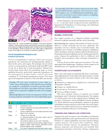
Figure 24.2 Vulval intraepithelial neoplasia (VIN) lesion (Bowen’s Histologically, the vaginal wall consists of 3 layers: an outer
disease). There is hyperkeratosis, parakeratosis, acanthosis, koilocytosis fibrous, a middle muscular and an inner epithelial. The
and presence of atypical anaplastic cells throughout the entire thickness muscular coat has a double layer of smooth muscle. The
of the epithelium. Photomicrograph on right under higher magnification
shows mitotic figures in the layers of squamous epithelium. epithelial layer consists of stratified squamous epithelium
which undergoes cytologic changes under hormonal stimuli.
Vulval Intraepithelial Neoplasia and Oestrogen increases its thickness such as during reproductive
Invasive Carcinoma years, whereas the epithelium is thin in childhood, and
atrophic after menopause when oestrogen stimulation is
Vulval intraepithelial neoplasia (VIN) and invasive minimal.
squamous cell carcinoma are morphologically similar to Primary diseases of the vagina are uncommon. The only CHAPTER 24
those in the cervix and vagina. The etiologic role of certain important clinicopathologic conditions which require to be
viruses in carcinogenesis, particularly high-risk HPV types described here are vaginitis and certain tumours.
16 and 18, in these sites is well documented. Mention has
already been made about the preceding stage of vulval VAGINITIS AND VULVOVAGINITIS
epithelial disorders, particularly squamous hyperplasia, in
the development of these lesions. Vulval carcinoma Since vulva and vagina are anatomically close to each other,
constitutes 3% of all female genital tract cancers. The usual often inflammation of one affects the other location. Certain
age for development of cancer or VIN is the 4th to 6th decade. other infections are quite common in the vulva and vagina
as follows:
Grossly, VIN and vulval carcinoma in early stage is a Bacterial e.g. streptococci, staphylococci, Escherichia coli,
‘white’ lesion (leukoplakia) while later the area develops Haemophilus vaginalis. The Female Genital Tract
an exophytic or endophytic (ulcerative) growth pattern.
The traditional VIN lesion, described as Bowen’s disease Fungal e.g. Candida albicans.
of the vulva, is generally a slightly elevated velvety plaque Protozoal e.g. Trichomonas vaginalis.
lesion. Viral e.g. Herpes simplex.
Microscopically, these lesions are squamous cell type with The most common causes of vaginitis are Candida
varying anaplasia and depth of invasion depending upon (moniliasis) and Trichomonas (trichomoniasis). The hyphae
of Candida can be seen in the vaginal smears. Similarly, the
protozoa, Trichomonas, can be identified in smears (Chapter
TABLE 24.1: FIGO Staging of Carcinoma of the Vulva. 11). These infections are particularly common in pregnant
and diabetic women and may involve both vulva and vagina.
Stage 0 Carcinoma in situ. However, the adult vaginal mucosa is relatively resistant to
Stage I Tumour confined to the vulva and/or perineum; 2 cm gonococcal infection because of its histology.
or less in diameter.
Stage II Tumour confined to the vulva and/or perineum; more
than 2 cm in diameter. TUMOURS AND TUMOUR-LIKE CONDITIONS
Stage III Tumour of any size with
(1) adjacent spread to the lower urethra and/ or Vaginal cysts such as Gartner’s duct (Wolffian) cyst lined by
vagina, or the anus, and/or glandular epithelium and vaginal inclusion cyst arising from
(2) unilateral regional lymph node metastasis. inclusion of vaginal epithelium are more common benign
Stage IVA Tumour invades any of the following—upper urethra, vaginal tumours and tumour-like conditions. Other
bladder mucosa, rectal mucosa, pelvic bone, and/or uncommon benign tumours are papillomas, fibromas,
bilateral regional node metastasis. lipomas, angiomas and leiomyomas and resemble their
Stage IVB Any distant metastasis including pelvic lymph nodes. counterparts elsewhere in the body. Primary malignancies