
Page 735 - Textbook of Pathology, 6th Edition
P. 735
719
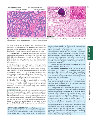
Figure 23.15 Carcinoma of the prostate. The field shows microacini of small malignant cells infiltrating the prostatic stroma. Inset in the
photomicrograph shows perineural invasion by prostatic adenocarcinoma.
cancer. It is uncommon in Japanese and Chinese, while the prostate is firm and fibrous. Cut section is homogeneous
prevalence is high in Americans. African Americans have a and contains irregular yellowish areas.
markedly higher incidence as compared to whites which may Microscopically, 4 histologic types are described—
be related to genetic variation in androgen receptor gene. adenocarcinoma, transitional cell carcinoma, squamous
3. Environmental influences. Some common environ- cell carcinoma and undifferentiated carcinoma. However, CHAPTER 23
mental factors and carcinogens have been identified with adenocarcinoma is the most common type found in 96% of
high risk to development of prostatic cancer. These include cases and is the one generally referred to as carcinoma of
high dietary fat, and exposure to polycyclic aromatic the prostate. The other three histologic types are rare and
hydrocarbons. Flavonoids, antioxidants and selenium may resemble in morphology with similar malignant tumours
reduce the risk. elsewhere in the body.
4. Nodular hyperplasia. Though nodular prostatic The histologic characteristics of adenocarcinoma of the
hyperplasia has been suggested by some as precursor for prostate are as under (Fig. 23.15):
development of prostatic cancer, it is considered unlikely. 1. Architectural disturbance. In contrast to convoluted
Most prostatic cancers develop in the periphery of the gland appearance of the glands seen in normal and hyperplastic
while BEP occurs in the periurethral part of the gland. Any prostate, there is loss of intra-acinar papillary
concomitant occurrence of the two diseases may be convolutions. The groups of acini are either closely packed
considered as aging process. Approximately 15-20% of in back-to-back arrangement without intervening stroma
nodular hyperplastic prostates harbour carcinoma. or are haphazardly distributed.
5. Heredity. The possibility of genetic basis of prostatic 2. Stroma. Normally, fibromuscular sling surrounds the
cancer has been suggested by the observations of familial acini, whereas malignant acini have little or no stroma The Male Reproductive System and Prostate
clustering and 2-fold higher frequency in first-degree between them. The tumour cells may penetrate and
relatives. Prostatic cancer susceptibility gene has been replace the fibromuscular stroma.
identified in familial cases. 3. Gland pattern. Most frequently, the glands in well-
HISTOGENESIS. Histogenesis of prostatic adenocarcinoma differentiated prostatic adenocarcinoma are small or
has been documented as a mutistep process arising from medium-sized, lined by a single layer of cuboidal or low
premalignant stage of prostatic intraepithelial neoplasia (PIN). columnar cells. Moderately-differentiated tumours have
PIN refers to multiple foci of cytologically atypical luminal cribriform or fenestrated glandular appearance. Poorly-
cells overlying diminished number of basal cells in prostatic differentiated tumours have little or no glandular arrange-
ducts and is a forerunner of invasive prostatic carcinoma. ment but instead show solid or trabecular pattern.
Based on cytologic atypia, PIN may be low grade to high 4. Tumour cells. In many cases, the individual tumour
grade. PIN of high-grade progresses to prostatic adeno- cells in prostatic carcinoma do not show usual
carcinoma. morphologic features of malignancy. The tumour cells
may be clear, dark and eosinophilic cells. Clear cells have
MORPHOLOGIC FEATURES. Grossly, the prostate may foamy cytoplasm, dark cells have homogeneous basophilic
be enlarged, normal in size or smaller than normal. In 95% cytoplasm, and eosinophilic cells have granular cytoplasm.
of cases, prostatic carcinoma is located in the peripheral The cells may show varying degree of anaplasia and
zone, especially in the posterior lobe. The malignant nuclear atypia but is generally slight.