
Page 765 - Textbook of Pathology, 6th Edition
P. 765
749
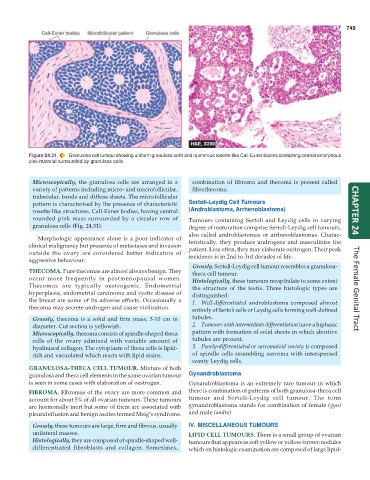
Figure 24.31 Granulosa cell tumour showing uniform granulosa cells and numerous rosette-like Call-Exner bodies containing central amorphous
pink material surrounded by granulosa cells.
Microscopically, the granulosa cells are arranged in a combination of fibroma and thecoma is present called
variety of patterns including micro- and macrofollicular, fibrothecoma.
trabecular, bands and diffuse sheets. The microfollicular
pattern is characterised by the presence of characteristic Sertoli-Leydig Cell Tumours
rosette-like structures, Call-Exner bodies, having central (Androblastoma, Arrhenoblastoma) CHAPTER 24
rounded pink mass surrounded by a circular row of Tumours containing Sertoli and Leydig cells in varying
granulosa cells (Fig. 24.31). degree of maturation comprise Sertoli-Leydig cell tumours,
also called androblastomas or arrhenoblastomas. Charac-
Morphologic appearance alone is a poor indicator of
clinical malignancy but presence of metastases and invasion teristically, they produce androgens and masculinise the
patient. Less often, they may elaborate oestrogen. Their peak
outside the ovary are considered better indicators of incidence is in 2nd to 3rd decades of life.
aggressive behaviour.
Grossly, Sertoli-Leydig cell tumour resembles a granulosa-
THECOMA. Pure thecomas are almost always benign. They theca cell tumour.
occur more frequently in postmenopausal women. Histologically, these tumours recapitulate to some extent
Thecomas are typically oestrogenic. Endometrial the structure of the testis. Three histologic types are
hyperplasia, endometrial carcinoma and cystic disease of distinguished: The Female Genital Tract
the breast are some of its adverse effects. Occasionally a 1. Well-differentiated androblastoma composed almost
thecoma may secrete androgen and cause virilisation.
entirely of Sertoli cells or Leydig cells forming well-defined
Grossly, thecoma is a solid and firm mass, 5-10 cm in tubules.
diameter. Cut section is yellowish. 2. Tumours with intermediate differentiation have a biphasic
Microscopically, thecoma consists of spindle-shaped theca pattern with formation of solid sheets in which abortive
cells of the ovary admixed with variable amount of tubules are present.
hyalinised collagen. The cytoplasm of theca cells is lipid- 3. Poorly-differentiated or sarcomatoid variety is composed
rich and vacuolated which reacts with lipid stains. of spindle cells resembling sarcoma with interspersed
scanty Leydig cells.
GRANULOSA-THECA CELL TUMOUR. Mixture of both
granulosa and theca cell elements in the same ovarian tumour Gynandroblastoma
is seen in some cases with elaboration of oestrogen. Gynandroblastoma is an extremely rare tumour in which
FIBROMA. Fibromas of the ovary are more common and there is combination of patterns of both granulosa-theca cell
account for about 5% of all ovarian tumours. These tumours tumour and Sertoli-Leydig cell tumour. The term
are hormonally inert but some of them are associated with gynandroblastoma stands for combination of female (gyn)
pleural effusion and benign ascites termed Meig’s syndrome. and male (andro).
Grossly, these tumours are large, firm and fibrous, usually IV. MISCELLANEOUS TUMOURS
unilateral masses. LIPID CELL TUMOURS. There is a small group of ovarian
Histologically, they are composed of spindle-shaped well- tumours that appears as soft yellow or yellow-brown nodules
differentiated fibroblasts and collagen. Sometimes, which on histologic examination are composed of large lipid-