
Page 763 - Textbook of Pathology, 6th Edition
P. 763
747
Figure 24.27 Benign cystic teratoma. Microscopy shows characteristic lining of the cyst wall by epidermis and its appendages. Islands of
mature cartilage are also seen.
having an embryonic appearance. Immature tissue patients with dysgerminoma have elevated hCG level in the
elements may differentiate towards cartilage, bone, plasma. All dysgerminomas are malignant and are extremely
glandular structures, neural tissue etc, and are distributed radiosensitive.
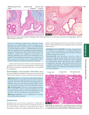
in spindle-shaped myxoid or undifferentiated sarcoma MORPHOLOGIC FEATURES. Grossly, dysgerminoma
cells. An important factor in grading and determining the is a solid mass of variable size. Cut section of the tumour CHAPTER 24
prognosis of immature teratoma is the relative amount of is grey-white to pink, lobulated, soft and fleshy with foci
immature neural tissue. Immature neural tissue can of haemorrhages and necrosis.
undergo maturation even at the site of metastases over a Histologically, their structure is similar to that of
period of years. Immature teratoma may contain areas of seminoma of the testis (Fig. 24.28). The tumour cells are
other germ cell tumours such as endodermal sinus arranged in diffuse sheets, islands and cords separated
tumour, embryonal carcinoma and choriocarcinoma.
by scanty fibrous stroma. The tumour cells are uniform
Grade I tumours having relatively mature elements and in appearance and large, with vesicular nuclei and clear
confined to the ovary have a good prognosis, whereas grade cytoplasm rich in glycogen. The fibrous stroma generally
III immature teratomas with metastases have an extremely contains lymphocytic infiltrate and sometimes may have
poor prognosis. sarcoid granulomas. The Female Genital Tract
MONODERMAL (SPECIALISED) TERATOMA. Mono-
dermal or highly specialised teratomas are rare and include
2 important examples—struma ovarii and carcinoid tumour.
Struma ovarii. It is a teratoma composed exclusively
of thyroid tissue, recognisable grossly as well as micros-
copically. Most often, the tumour has the appearance of a
follicular adenoma of the thyroid. Rarely, struma ovarii
may be hyperfunctioning and produce hyperthyroidism.
Carcinoid tumour. This is an ovarian teratoma arising
from argentaffin cells of intestinal epithelium in the
teratoma. Ovarian carcinoid may also hyperfunction and
produce 5-HT and consequent carcinoid syndrome.
Struma-carcinoid is a rare combination of struma
ovarii and ovarian carcinoid.
Dysgerminoma
Dysgerminoma is an ovarian counterpart of seminoma of
the testes (page 709). Dysgerminomas comprise about 2% of Figure 24.28 Dysgerminoma. The histologic appearance is identical
all ovarian cancers. They occur most commonly in 2nd to to that of seminoma of the testis. Masses of large uniform tumour cells
3rd decades. About 10% of them are bilateral. About 10% of are separated by scanty fibrous stroma that is infiltrated by lymphocytes.