
Page 766 - Textbook of Pathology, 6th Edition
P. 766
750
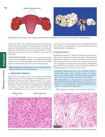
Figure 24.32 Krukenberg tumour showing characteristic bilateral metastatic ovarian cancer (arrow) having firm white appearance.
laden cells. These cells resemble Leydig, lutein and adrenal carcinomas of the breast, genital tract, gastrointestinal tract
cortical cells. The examples of these tumours are: hilus cell (e.g. stomach, colon appendix, pancreas, biliary tract) and
tumours, adrenal rest tumours and luteomas. These tumours haematopoietic malignancies.
elaborate steroid hormones and are responsible for various
endocrine dysfunctions such as Cushing’s syndrome and Krukenberg Tumour
virilisation.
Krukenberg tumour is a distinctive bilateral tumour meta-
GONADOBLASTOMA. This is a rare tumour occurring static to the ovaries by transcoelomic spread. The tumour is
exclusively in dysgenetic gonads, more often in phenotypic generally secondary to a gastric carcinoma (page 557) but
females and in hermaphrodites. Dysfunctions include other primary sites where mucinous carcinomas occur (e.g.
virilism, amenorrhoea and abnormal external genitalia. colon, appendix and breast) may also produce Krukenberg
tumour in the ovary. Rarely, a tumour having the pattern of
Microscopically, gonadoblastoma is composed of mixture Krukenberg tumour is primary in the ovary.
SECTION III
of germ cell and sex cord components.
Grossly, Krukenberg tumour forms rounded or kidney-
V. METASTATIC TUMOURS shaped firm large masses in both ovaries. Cut section
shows grey-white to yellow firm fleshy tumour and may
About 10% of ovarian cancers are secondary carcinomas. have areas of haemorrhage and necrosis (Fig. 24.32).
Metastasis may occur by lymphatic or haematogenous route
but direct extension from adjacent organs (e.g. uterus, Microscopically, it is characterised by the presence of
fallopian tube and sigmoid colon) too occurs frequently. mucus-filled signet ring cells which may lie singly or in
Bilaterality of the tumour is the most helpful clue to diagnosis clusters. It is accompanied by sarcoma-like cellular prolife-
of metastatic tumour. Most common primary sites from ration of ovarian stroma (Fig. 24.33).
where metastases to the ovaries are encountered are: FIGO staging of ovarian cancer is given in Table 24.7.
Systemic Pathology
Figure 24.33 Krukenberg tumour. Histologic features include mucin-filled signet-ring cells and richly cellular proliferation of the ovarian stroma.