
Page 764 - Textbook of Pathology, 6th Edition
P. 764
748 Endodermal Sinus (Yolk Sac) Tumour
Endodermal sinus tumour or yolk sac tumour is the second
most common germ cell tumour occurring most frequently
in children and young women. More often, endodermal
sinus tumour is found in combination with other germ cell
tumours rather than in pure form. The tumour is rich in
alphafetoprotein (AFP) and α-1-antitrypsin. The tumour is
usually unilateral but may metastasise to the other ovary.
It is a highly aggressive and rapidly growing tumour.
MORPHOLOGIC FEATURES. Grossly, the tumour is
generally solid with areas of cystic degeneration.
Histologically, like its testicular counterpart, the endo-
dermal sinus tumour is characterised by the presence of
papillary projections having a central blood vessel with
perivascular layer of anaplastic embryonal germ cells.
Such structures resemble the endodermal sinuses of the
rat placenta (Schiller-Duval body) from which the tumour
derives its name. It is common to find intracellular and
extracelluar PAS-positive hyaline globules which are
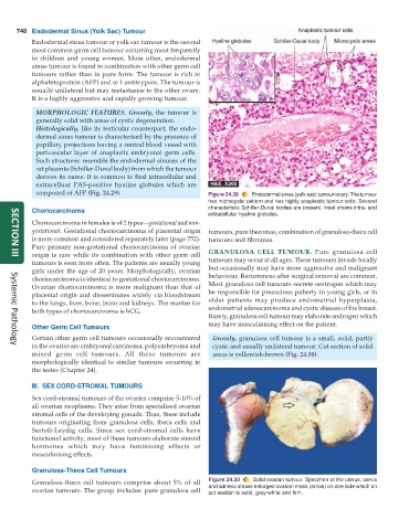
composed of AFP (Fig. 24.29). Figure 24.29 Endodermal sinus (yolk sac) tumour ovary. The tumour
has microcystic pattern and has highly anaplastic tumour cells. Several
Choriocarcinoma characteristic Schiller-Duval bodies are present. Inset shows intra- and
extracellular hyaline globules.
Choriocarcinoma in females is of 2 types—gestational and non-
gestational. Gestational choriocarcinoma of placental origin tumours, pure thecomas, combination of granulosa-theca cell
is more common and considered separately later (page 752). tumours and fibromas.
Pure primary non-gestational choriocarcinoma of ovarian
origin is rare while its combination with other germ cell GRANULOSA CELL TUMOUR. Pure granulosa cell
SECTION III
tumours is seen more often. The patients are usually young tumours may occur at all ages. These tumours invade locally
girls under the age of 20 years. Morphologically, ovarian but occasionally may have more aggressive and malignant
choriocarcinoma is identical to gestational choriocarcinoma. behaviour. Recurrences after surgical removal are common.
Ovarian choriocarcinoma is more malignant than that of Most granulosa cell tumours secrete oestrogen which may
placental origin and disseminates widely via bloodstream be responsible for precocious puberty in young girls, or in
to the lungs, liver, bone, brain and kidneys. The marker for older patients may produce endometrial hyperplasia,
both types of choriocarcinoma is hCG. endometrial adenocarcinoma and cystic disease of the breast.
Rarely, granulosa cell tumour may elaborate androgen which
may have masculinising effect on the patient.
Other Germ Cell Tumours
Certain other germ cell tumours occasionally encountered Grossly, granulosa cell tumour is a small, solid, partly
in the ovaries are embryonal carcinoma, polyembryoma and cystic and usually unilateral tumour. Cut section of solid
Systemic Pathology
mixed germ cell tumours. All these tumours are areas is yellowish-brown (Fig. 24.30).
morphologically identical to similar tumours occurring in
the testes (Chapter 24).
III. SEX CORD-STROMAL TUMOURS
Sex cord-stromal tumours of the ovaries comprise 5-10% of
all ovarian neoplasms. They arise from specialised ovarian
stromal cells of the developing gonads. Thus, these include
tumours originating from granulosa cells, theca cells and
Sertoli-Leydig cells. Since sex cord-stromal cells have
functional activity, most of these tumours elaborate steroid
hormones which may have feminising effects or
masculinising effects.
Granulosa-Theca Cell Tumours
Granulosa-theca cell tumours comprise about 5% of all Figure 24.30 Solid ovarian tumour. Specimen of the uterus, cervix
ovarian tumours. The group includes: pure granulosa cell and adnexa shows enlarged ovarian mass (arrow) on one side which on
cut section is solid, grey-white and firm.