
Page 778 - Textbook of Pathology, 6th Edition
P. 778
762
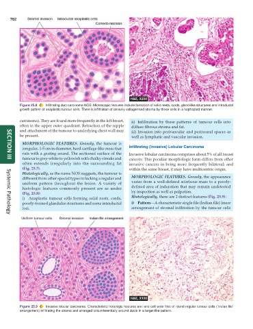
Figure 25.8 Infiltrating duct carcinoma-NOS. Microscopic features include formation of solid nests, cords, gland-like structures and intraductal
growth pattern of anaplastic tumour cells. There is infiltration of densely collagenised stroma by these cells in a haphazard manner.
carcinoma). They are found more frequently in the left breast, ii) Infiltration by these patterns of tumour cells into
often in the upper outer quadrant. Retraction of the nipple diffuse fibrous stroma and fat.
and attachment of the tumour to underlying chest wall may iii) Invasion into perivascular and perineural spaces as
be present. well as lymphatic and vascular invasion.
MORPHOLOGIC FEATURES. Grossly, the tumour is
irregular, 1-5 cm in diameter, hard cartilage-like mass that Infiltrating (Invasive) Lobular Carcinoma
cuts with a grating sound. The sectioned surface of the Invasive lobular carcinoma comprises about 5% of all breast
tumour is grey-white to yellowish with chalky streaks and cancers. This peculiar morphologic form differs from other
SECTION III
often extends irregularly into the surrounding fat invasive cancers in being more frequently bilateral; and
(Fig. 25.7). within the same breast, it may have multicentric origin.
Histologically, as the name NOS suggests, the tumour is
different from other special types in lacking a regular and MORPHOLOGIC FEATURES. Grossly, the appearance
uniform pattern throughout the lesion. A variety of varies from a well-defined scirrhous mass to a poorly-
histologic features commonly present are as under defined area of induration that may remain undetected
(Fig. 25.8): by inspection as well as palpation.
i) Anaplastic tumour cells forming solid nests, cords, Histologically, there are 2 distinct features (Fig. 25.9):
poorly-formed glandular structures and some intraductal i) Pattern—A characteristic single file (Indian file) linear
foci. arrangement of stromal infiltration by the tumour cells
Systemic Pathology
Figure 25.9 Invasive lobular carcinoma. Characteristic histologic features are: one cell wide files of round regular tumour cells (‘Indian file’
arrangement) infiltrating the stroma and arranged circumferentially around ducts in a target-like pattern.