
Page 779 - Textbook of Pathology, 6th Edition
P. 779
with very little tendency to gland formation is seen. Colloid (Mucinous) Carcinoma 763
Infiltrating cells may be arranged concentrically around This is an uncommon pattern of breast cancer occurring more
ducts in a target-like pattern. frequently in older women and is slow-growing. Colloid
ii) Tumour cytology—Individual tumour cells resemble carcinoma has better prognosis than the usual infiltrating
cells of in situ lobular carcinoma. They are round and duct carcinoma.
regular with very little pleomorphism and infrequent MORPHOLOGIC FEATURES. Grossly, the tumour is
mitoses. Some tumours may show signet-ring cells usually a soft and gelatinous mass with well-demarcated
distended with cytoplasmic mucin.
borders.
Histologically, colloid carcinoma contains large amount
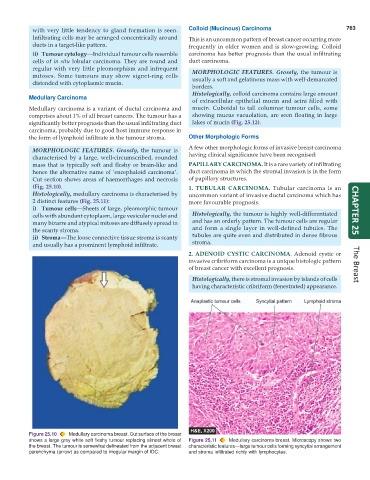
Medullary Carcinoma
of extracellular epithelial mucin and acini filled with
Medullary carcinoma is a variant of ductal carcinoma and mucin. Cuboidal to tall columnar tumour cells, some
comprises about 1% of all breast cancers. The tumour has a showing mucus vacuolation, are seen floating in large
significantly better prognosis than the usual infiltrating duct lakes of mucin (Fig. 25.12).
carcinoma, probably due to good host immune response in
the form of lymphoid infiltrate in the tumour stroma. Other Morphologic Forms
A few other morphologic forms of invasive breast carcinoma
MORPHOLOGIC FEATURES. Grossly, the tumour is
characterised by a large, well-circumscribed, rounded having clinical significance have been recognised:
mass that is typically soft and fleshy or brain-like and PAPILLARY CARCINOMA. It is a rare variety of infiltrating
hence the alternative name of ‘encephaloid carcinoma’. duct carcinoma in which the stromal invasion is in the form
Cut section shows areas of haemorrhages and necrosis of papillary structures.
(Fig. 25.10). 1. TUBULAR CARCINOMA. Tubular carcinoma is an
Histologically, medullary carcinoma is characterised by uncommon variant of invasive ductal carcinoma which has
2 distinct features (Fig. 25.11): more favourable prognosis.
i) Tumour cells—Sheets of large, pleomorphic tumour CHAPTER 25
cells with abundant cytoplasm, large vesicular nuclei and Histologically, the tumour is highly well-differentiated
many bizarre and atypical mitoses are diffusely spread in and has an orderly pattern. The tumour cells are regular
the scanty stroma. and form a single layer in well-defined tubules. The
ii) Stroma—The loose connective tissue stroma is scanty tubules are quite even and distributed in dense fibrous
and usually has a prominent lymphoid infiltrate. stroma.
2. ADENOID CYSTIC CARCINOMA. Adenoid cystic or
invasive cribriform carcinoma is a unique histologic pattern
of breast cancer with excellent prognosis. The Breast
Histologically, there is stromal invasion by islands of cells
having characteristic cribriform (fenestrated) appearance.
Figure 25.10 Medullary carcinoma breast. Cut surface of the breast
shows a large grey white soft fleshy tumour replacing almost whole of Figure 25.11 Medullary carcinoma breast. Microscopy shows two
the breast. The tumour is somewhat delineated from the adjacent breast characteristic features—large tumour cells forming syncytial arrangement
parenchyma (arrow) as compared to irregular margin of IDC. and stroma infiltrated richly with lymphocytes.