
Page 805 - Textbook of Pathology, 6th Edition
P. 805
789
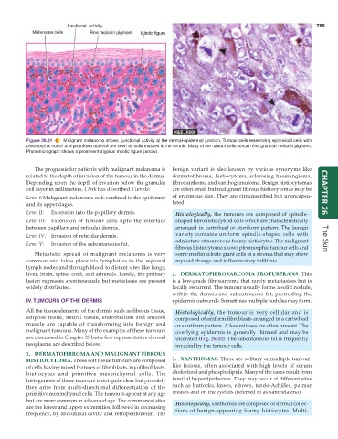
Figure 26.31 Malignant melanoma shows junctional activity at the dermal-epidermal junction. Tumour cells resembling epithelioid cells with
pleomorphic nuclei and prominent nucleoli are seen as solid masses in the dermis. Many of the tumour cells contain fine granular melanin pigment.
Photomicrograph shows a prominent atypical mitotic figure (arrow).
The prognosis for patients with malignant melanoma is benign variant is also known by various synonyms like
related to the depth of invasion of the tumour in the dermis. dermatofibroma, histiocytoma, sclerosing haemangioma,
Depending upon the depth of invasion below the granular fibroxanthoma and xanthogranuloma. Benign histiocytomas
cell layer in millimeters, Clark has described 5 levels: are often small but malignant fibrous histiocytomas may be
Level I: Malignant melanoma cells confined to the epidermis of enormous size. They are circumscribed but unencapsu- CHAPTER 26
and its appendages. lated.
Level II: Extension into the papillary dermis. Histologically, the tumours are composed of spindle-
Level III: Extension of tumour cells upto the interface shaped fibrohistiocytoid cells which are characteristically
between papillary and reticular dermis. arranged in cartwheel or storiform pattern. The benign
Level IV: Invasion of reticular dermis. variety contains uniform spindle-shaped cells with
admixture of numerous foamy histiocytes. The malignant The Skin
Level V: Invasion of the subcutaneous fat.
fibrous histiocytoma shows pleomorphic tumour cells and
Metastatic spread of malignant melanoma is very some multinucleate giant cells in a stroma that may show
common and takes place via lymphatics to the regional myxoid change and inflammatory infiltrate.
lymph nodes and through blood to distant sites like lungs,
liver, brain, spinal cord, and adrenals. Rarely, the primary 2. DERMATOFIBROSARCOMA PROTUBERANS. This
lesion regresses spontaneously but metastases are present is a low-grade fibrosarcoma that rarely metastasises but is
widely distributed. locally recurrent. The tumour usually forms a solid nodule,
within the dermis and subcutaneous fat, protruding the
IV.TUMOURS OF THE DERMIS epidermis outwards. Sometimes multiple nodules may form.
All the tissue elements of the dermis such as fibrous tissue, Histologically, the tumour is very cellular and is
adipose tissue, neural tissue, endothelium and smooth composed of uniform fibroblasts arranged in a cartwheel
muscle are capable of transforming into benign and or storiform pattern. A few mitoses are often present. The
malignant tumours. Many of the examples of these tumours overlying epidermis is generally thinned and may be
are discussed in Chapter 29 but a few representative dermal ulcerated (Fig. 26.32). The subcutaneous fat is frequently
neoplasms are described below. invaded by the tumour cells.
1. DERMATOFIBROMA AND MALIGNANT FIBROUS
HISTIOCYTOMA. These soft tissue tumours are composed 3. XANTHOMAS. These are solitary or multiple tumour-
of cells having mixed features of fibroblasts, myofibroblasts, like lesions, often associated with high levels of serum
histiocytes and primitive mesenchymal cells. The cholesterol and phospholipids. Many of the cases result from
histogenesis of these tumours is not quite clear but probably familial hyperlipidaemia. They may occur at different sites
they arise from multi-directional differentiation of the such as buttocks, knees, elbows, tendo-Achilles, palmar
primitive mesenchymal cells. The tumours appear at any age creases and on the eyelids (referred to as xanthelasma).
but are more common in advanced age. The commonest sites Histologically, xanthomas are composed of dermal collec-
are the lower and upper extremities, followed in decreasing tions of benign-appearing foamy histiocytes. Multi-
frequency, by abdominal cavity and retroperitoneum. The