
Page 800 - Textbook of Pathology, 6th Edition
P. 800
784
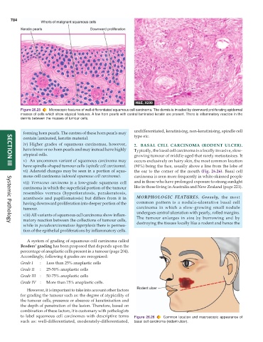
Figure 26.25 Microscopic features of well-differentiated squamous cell carcinoma. The dermis is invaded by downward proliferating epidermal
masses of cells which show atypical features. A few horn pearls with central laminated keratin are present. There is inflammatory reaction in the
dermis between the masses of tumour cells.
undifferentiated, keratinising, non-keratinising, spindle cell
forming horn pearls. The centres of these horn pearls may
contain laminated, keratin material. type etc.
iv) Higher grades of squamous carcinomas, however, 2. BASAL CELL CARCINOMA (RODENT ULCER).
have fewer or no horn pearls and may instead have highly Typically, the basal cell carcinoma is a locally invasive, slow-
atypical cells. growing tumour of middle-aged that rarely metastasises. It
v) An uncommon variant of squamous carcinoma may occurs exclusively on hairy skin, the most common location
have spindle-shaped tumour cells (spindle cell carcinoma). (90%) being the face, usually above a line from the lobe of
SECTION III
vi) Adenoid changes may be seen in a portion of squa- the ear to the corner of the mouth (Fig. 26.26). Basal cell
mous cell carcinoma (adenoid squamous cell carcinoma). carcinoma is seen more frequently in white-skinned people
vii) Verrucous carcinoma is a low-grade squamous cell and in those who have prolonged exposure to strong sunlight
carcinoma in which the superficial portion of the tumour like in those living in Australia and New Zealand (page 221).
resembles verruca (hyperkeratosis, parakeratosis,
acanthosis and papillomatosis) but differs from it in MORPHOLOGIC FEATURES. Grossly, the most
having downward proliferation into deeper portion of the common pattern is a nodulo-ulcerative basal cell
tumour. carcinoma in which a slow-growing small nodule
undergoes central ulceration with pearly, rolled margins.
viii) All variants of squamous cell carcinoma show inflam-
matory reaction between the collections of tumour cells, The tumour enlarges in size by burrowing and by
while in pseudocarcinomatous hyperplasia there is permea- destroying the tissues locally like a rodent and hence the
Systemic Pathology
tion of the epithelial proliferations by inflammatory cells.
A system of grading of squamous cell carcinoma called
Broders’ grading has been proposed that depends upon the
percentage of anaplastic cells present in a tumour (page 204).
Accordingly, following 4 grades are recognised:
Grade I : Less than 25% anaplastic cells
Grade II : 25-50% anaplastic cells
Grade III : 50-75% anaplastic cells
Grade IV : More than 75% anaplastic cells.
However, it is important to take into account other factors
for grading the tumour such as: the degree of atypicality of
the tumour cells, presence or absence of keratinisation and
the depth of penetration of the lesion. Therefore, based on
combination of these factors, it is customary with pathologists
to label squamous cell carcinomas with descriptive terms Figure 26.26 Common location and macroscopic appearance of
such as: well-differentiated, moderately-differentiated, basal cell carcinoma (rodent ulcer).