
Page 801 - Textbook of Pathology, 6th Edition
P. 801
785
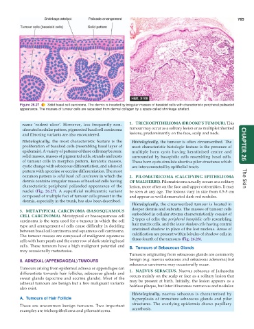
Figure 26.27 Solid basal cell carcinoma. The dermis is invaded by irregular masses of basaloid cells with characteristic peripheral palisaded
appearance. The masses of tumour cells are separated from dermal collagen by a space called shrinkage artefact.
name ‘rodent ulcer’. However, less frequently non- 1. TRICHOEPITHELIOMA (BROOKE’S TUMOUR). This
ulcerated nodular pattern, pigmented basal cell carcinoma tumour may occur as a solitary lesion or as multiple inherited
and fibrosing variants are also encountered. lesions, predominantly on the face, scalp and neck.
Histologically, the most characteristic feature is the Histologically, the tumour is often circumscribed. The
proliferation of basaloid cells (resembling basal layer of most characteristic histologic feature is the presence of CHAPTER 26
epidermis). A variety of patterns of these cells may be seen: multiple horn cysts having keratinised centre and
solid masses, masses of pigmented cells, strands and nests surrounded by basophilic cells resembling basal cells.
of tumour cells in morphea pattern, keratotic masses, These horn cysts simulate abortive pilar structures which
cystic change with sebaceous differentiation, and adenoid are interconnected by epithelial tracts.
pattern with apocrine or eccrine differentiation. The most
common pattern is solid basal cell carcinoma in which the 2. PILOMATRICOMA (CALCIFYING EPITHELIOMA
dermis contains irregular masses of basaloid cells having OF MALHERBE). Pilomatricoma usually occurs as a solitary The Skin
characteristic peripheral palisaded appearance of the lesion, more often on the face and upper extremities. It may
nuclei (Fig. 26.27). A superficial multicentric variant be seen at any age. The lesions vary in size from 0.5-5 cm
composed of multiple foci of tumour cells present in the and appear as well-demarcated dark red nodules.
dermis, especially in the trunk, has also been described.
Histologically, the circumscribed tumour is located in
deeper dermis and subcutis. The masses of tumour cells
3. METATYPICAL CARCINOMA (BASOSQUAMOUS
CELL CARCINOMA). Metatypical or basosquamous cell embedded in cellular stroma characteristically consist of
carcinoma is the term used for a tumour in which the cell 2 types of cells: the peripheral basophilic cells resembling
type and arrangement of cells cause difficulty in deciding hair matrix cells, and the inner shadow cells having central
between basal cell carcinoma and squamous cell carcinoma. unstained shadow in place of the lost nucleus. Areas of
The tumour masses are composed of malignant squamous calcification are present within lobules of shadow cells in
cells with horn pearls and the outer row of dark-staining basal three-fourth of the tumours (Fig. 26.28).
cells. These tumours have a high malignant potential and B. Tumours of Sebaceous Glands
may occasionally metastasise.
Tumours originating from sebaceous glands are commonly
II. ADNEXAL (APPENDAGEAL) TUMOURS benign (e.g. naevus sebaceus and sebaceous adenoma) but
sebaceous carcinoma may occasionally occur.
Tumours arising from epidermal adnexa or appendages can 1. NAEVUS SEBACEUS. Naevus sebaceus of Jadassohn
differentiate towards hair follicles, sebaceous glands and occurs mainly on the scalp or face as a solitary lesion that
sweat glands (apocrine and eccrine glands). Most of the may be present at birth. Initially, the lesion appears as a
adnexal tumours are benign but a few malignant variants hairless plaque, but later it becomes verrucous and nodular.
also exist.
Histologically, naevus sebaceus is characterised by
A. Tumours of Hair Follicle hyperplasia of immature sebaceous glands and pilar
structures. The overlying epidermis shows papillary
These are uncommon benign tumours. Two important
examples are trichoepithelioma and pilomatricoma. acanthosis.