
Page 803 - Textbook of Pathology, 6th Edition
P. 803
III. MELANOCYTIC TUMOURS 787
Melanocytic tumours may arise from one of the three cell
types: naevus cells, epidermal melanocytes and dermal
melanocytes.
Benign tumours originating from naevus cells are called
naevocellular naevi.
The examples of benign tumours arising from epidermal
melanocytes are lentigo, freckles, pigmentation associated with
Albright’s syndrome and cafe-au-lait spots of neuro-
fibromatosis (page 838).
Benign tumours derived from dermal melanocytes are
Mongolian spots, naevi of Ota and of Ito and the blue naevus.
Malignant melanoma is the malignant counterpart of
melanocytic tumours.
The important examples amongst these are described
below.
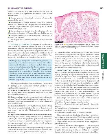
1. NAEVOCELLULAR NAEVI. Pigmented naevi or moles Figure 26.29 Intradermal naevus showing nests of naevus cells
are extremely common lesions on the skin of most which are typically uniform and present in the dermis. Melanin pigment
individuals. They are often flat or slightly elevated lesions; in naevus cells is coarse and irregular.
rarely they may be papillomatous or pedunculated. Most
naevi appear in adolescence and in early adulthood due to vii) Dysplastic naevi are certain atypical naevi which have
hormonal influence but rarely may be present at birth. They increased risk of progression to malignant melanoma. These
are mostly tan to brown and less than 1 cm in size. lesions are larger than the usual acquired naevi, are often
multiple, and appear as flat macules to slightly elevated
Histologically, irrespective of the histologic types, all plaques with irregular borders and variable pigmentation. CHAPTER 26
naevocellular naevi are composed of ‘naevus cells’ which Many of the cases are familial and inheritable. Dysplastic
are actually identical to melanocytes but differ from naevi have melanocytic proliferation at the epidermo-dermal
melanocytes in being arranged in clusters or nests. Naevus junction with some cytologic atypia.
cells are cuboidal or oval in shape with homogeneous
cytoplasm and contain large round or oval nucleus. 2. MALIGNANT MELANOMA. Malignant melanoma or
Melanin pigment is abundant in the naevus cells present melanocarcinoma arising from melanocytes is one of the most
in the lower epidermis and upper dermis, but the cells in rapidly spreading malignant tumour of the skin that can The Skin
the mid-dermis and lower dermis hardly contain any occur at all ages but is rare before puberty. The tumour
melanin (Fig. 26.29). spreads locally as well as to distant sites by lymphatics and
by blood. The etiology is unknown but there is role of
The important histological variants of naevi are as under: excessive exposure of white skin to sunlight e.g. higher
i) Lentigo is the replacement of the basal layer of the incidence in New Zealand and Australia where sun exposure
epidermis by melanocytes. in high. Besides the skin, melanomas may occur at various
ii) Junctional naevus is the one in which the naevus cells other sites such as oral and anogenital mucosa, oesophagus,
conjunctiva, orbit (page 512) and leptomeninges. The
lie at the epidermal-dermal junction. The naevus cells form
well-circumscribed nests. common sites on the skin are the trunk (in men), legs (in
women); other locations are face, soles, palms and nail-beds.
iii) Compound naevus is the commonest type of pigmented Some high risk factors associated with increased
naevus. These lesions, in addition to the junctional activity incidence of malignant melanoma are as under:
as in junctional naevi, show nests of naevus cells in the dermis i) Persistent change in appearance of a mole.
to a variable depth. ii) Presence of pre-existing naevus (especially dysplastic
iv) Intradermal naevus shows slight or no junctional activity. naevus).
The lesion is mainly located in the upper dermis as nests iii) Family history of melanoma in a patient of atypical mole.
and cords of naevus cells. Multinucleate naevus cells are iii) Higher age of the patient.
common. iv) More than 50 moles 2 mm or more in diameter.
v) Spindle cell (epithelioid) naevus or juvenile melanoma Molecular studies in familial and hereditary cases have
is a compound naevus with junctional activity. The naevus revealed germline mutation in CDKN2A gene which encodes
cells are, however, elongated and epithelioid in appearance for cyclin-dependent kinase inhibitor, mutational loss of
which may or may not contain melanin. Juvenile melanoma PTEN gene and mutation in several other tumour suppressor
is important since it is frequently confused with malignant genes but not p53.
melanoma histologically. Clinically, melanoma often appears as a flat or slightly
vi) Blue naevus is characterised by dendritic spindle naevus elevated naevus which has variegated pigmentation,
cells rather than the usual rounded or cuboidal naevus cells. irregular borders and, of late, has undergone secondary
These cells are often quite rich in melanin pigment. changes of ulceration, bleeding and increase in size. Many