
Page 804 - Textbook of Pathology, 6th Edition
P. 804
788
TABLE 26.3: Distinguishing Features of Benign Mole and Malignant Melanoma.
Feature Benign Mole Malignant Melanoma
1. Clinical features
i) Symmetry Symmetrical A = Asymmetry
ii) Border Well-demarcated B = Border irregularity
iii) Colour Uniformly pigmented C = Colour change
iv) Diameter Small, less than 6 mm D = Diameter more than 6 mm
2. Common locations Skin of face, mucosa Skin; mucosa of nose, bowel, anal region
3. Histopathology
i) Architecture Nests of cells Various patterns: solid sheets, alveoli, nests, islands
ii) Cell morphology Uniform looking naevus cells Malignant cells, atypia, mitoses, nucleoli
iii) Melanin pigment Irregular, coarse clumps Fine granules, uniformly distributed
iv) Inflammation May or may not be present Often present
4. Spread Remains confined, poses cosmetic Haematogenous and/or lymphatic spread early
problem only
of the malignant melanomas, however, arise de novo rather epithelioid or spindle-shaped, the former being more
than from a pre-existing naevus. Malignant melanoma can common. The tumour cells have amphophilic cytoplasm
be differentiated from benign pigmented lesions by subtle and large, pleomorphic nuclei with conspicuous nucleoli.
features as summed up in Table 26.3; the dermatologists term Mitotic figures are often present and multinucleate giant
this as ABCD of melanoma (acronym for Asymmetry, Border cells may occur. These tumour cells may be arranged in
irregularity, Colour change and Diameter >6mm). various patterns such as solid masses, sheets, island,
alveoli etc.
MORPHOLOGIC FEATURES. Grossly, depending upon iii) Melanin. Melanin pigment may be present (melanotic)
the clinical course and prognosis, cutaneous malignant or absent (amelanotic melanoma) without any prognostic
melanomas are of the following 4 types: influence. The pigment, if present, tends to be in the form
SECTION III
i) Lentigo maligna melanoma. This often develops from of uniform fine granules (unlike the benign naevi in which
a pre-existing lentigo (a flat naevus characterised by coarse irregular clumps of melanin are present). At times,
replacement of basal layer of epidermis by naevus cells). there may be no evidence of melanin in H&E stained
It is essentially a malignant melanoma in situ. It is slow- sections but Fontana-Masson stain or dopa reaction reveals
growing and has good prognosis. melanin granules in the cytoplasm of tumour cells.
ii) Superficial spreading melanoma. This is a slightly Immunohistochemically, melnoma cells are positive for
elevated lesion with variegated colour and ulcerated HMB-45 (most specific), S-100 and Melan-A.
surface. It often develops from a superficial spreading iv) Inflammatory infiltrate. Some amount of inflam-
melanoma in situ (pagetoid melanoma) in 5 to 7 years. matory infiltrate is present in the invasive melanomas.
The prognosis is worse than for lentigo maligna Infrequently, partial spontaneous regression of the tumour
melanoma. occurs due to destructive effect of dense inflammatory
Systemic Pathology
infiltrate.
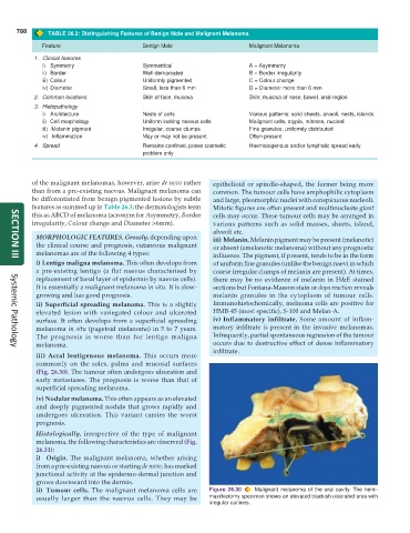
iii) Acral lentigenous melanoma. This occurs more
commonly on the soles, palms and mucosal surfaces
(Fig. 26.30). The tumour often undergoes ulceration and
early metastases. The prognosis is worse than that of
superficial spreading melanoma.
iv) Nodular melanoma. This often appears as an elevated
and deeply pigmented nodule that grows rapidly and
undergoes ulceration. This variant carries the worst
prognosis.
Histologically, irrespective of the type of malignant
melanoma, the following characteristics are observed (Fig.
26.31):
i) Origin. The malignant melanoma, whether arising
from a pre-existing naevus or starting de novo, has marked
junctional activity at the epidermo-dermal junction and
grows downward into the dermis.
ii) Tumour cells. The malignant melanoma cells are Figure 26.30 Malignant melanoma of the oral cavity. The hemi-
usually larger than the naevus cells. They may be maxillectomy specimen shows an elevated blackish ulcerated area with
irregular outlines.