
Page 856 - Textbook of Pathology, 6th Edition
P. 856
840
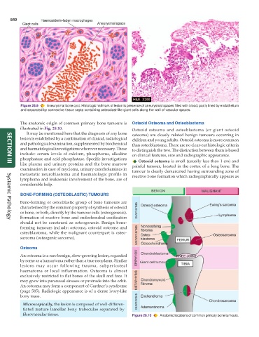
Figure 28.9 Aneurysmal bone cyst. Histologic hallmark of lesion is presence of aneurysmal spaces filled with blood, partly lined by endothelium
and separated by connective tissue septa containing osteoclast-like giant cells along the wall of vascular spaces.
The anatomic origin of common primary bone tumours is Osteoid Osteoma and Osteoblastoma
illustrated in Fig. 28.10. Osteoid osteoma and osteoblastoma (or giant osteoid
It may be mentioned here that the diagnosis of any bone osteoma) are closely related benign tumours occurring in
lesion is established by a combination of clinical, radiological children and young adults. Osteoid osteoma is more common
and pathological examination, supplemented by biochemical than osteoblastoma. There are no clear-cut histologic criteria
and haematological investigations wherever necessary. These to distinguish the two. The distinction between them is based
include: serum levels of calcium, phosphorus, alkaline on clinical features, size and radiographic appearance.
phosphatase and acid phosphatase. Specific investigations Osteoid osteoma is small (usually less than 1 cm) and
like plasma and urinary proteins and the bone marrow painful tumour, located in the cortex of a long bone. The
SECTION III
examination in case of myeloma, urinary catecholamines in tumour is clearly demarcated having surrounding zone of
metastatic neuroblastoma and haematologic profile in reactive bone formation which radiographically appears as
lymphoma and leukaemic involvement of the bone, are of
considerable help.
BONE-FORMING (OSTEOBLASTIC) TUMOURS
Bone-forming or osteoblastic group of bone tumours are
characterised by the common property of synthesis of osteoid
or bone, or both, directly by the tumour cells (osteogenesis).
Formation of reactive bone and endochondral ossification
Systemic Pathology
should not be construed as osteogenesis. Benign bone-
forming tumours include: osteoma, osteoid osteoma and
osteoblastoma, while the malignant counterpart is osteo-
sarcoma (osteogenic sarcoma).
Osteoma
An osteoma is a rare benign, slow-growing lesion, regarded
by some as a hamartoma rather than a true neoplasm. Similar
lesions may occur following trauma, subperiosteal
haematoma or local inflammation. Osteoma is almost
exclusively restricted to flat bones of the skull and face. It
may grow into paranasal sinuses or protrude into the orbit.
An osteoma may form a component of Gardner’s syndrome
(page 585). Radiologic appearance is of a dense ivory-like
bony mass.
Microscopically, the lesion is composed of well-differen-
tiated mature lamellar bony trabeculae separated by
fibrovascular tissue. Figure 28.10 Anatomic locations of common primary bone tumours.