
Page 855 - Textbook of Pathology, 6th Edition
P. 855
839
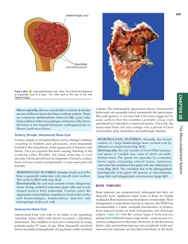
Figure 28.8 Aneurysmal bone cyst, ulna. The end of the long bone
is expanded due to a cyst. The inner wall of the cyst is tan and
haemorrhagic.
Microscopically, fibrous cortical defect consists of cellular column. The radiographic appearance shows characteristic CHAPTER 28
masses of fibrous tissue showing storiform pattern. There ballooned-out expansile lesion underneath the periosteum.
are numerous multinucleate osteoclast-like giant cells, The pathogenesis is not clear but it has been suggested by
haemosiderin-laden macrophages and foamy cells; hence some authors that the condition probably arises from
the lesion is also termed histiocytic xanthogranuloma or persistent local alteration in haemodynamics. Clinically, the
fibrous xanthoma of bone. aneurysmal bone cyst may enlarge over a period of years
and produce pain, tenderness and pathologic fracture.
Solitary (Simple, Unicameral) Bone Cyst
Solitary, simple or unicameral bone cyst is a benign condition MORPHOLOGIC FEATURES. Grossly, the lesion
occurring in children and adolescents, most frequently consists of a large haemorrhagic mass covered over by
located in the metaphyses at the upper end of humerus and thinned out reactive bone (Fig. 28.8).
femur. The cyst expands the bone causing thinning of the Histologically, the cyst consists of blood-filled aneurys- The Musculoskeletal System
overlying cortex. Possibly, the lesion arises due to local mal spaces of variable size, some of which are endo-
disorder of bone growth and development. Clinically, solitary thelium-lined. The spaces are separated by connective
bone cyst may remain asymptomatic or may cause pain and tissue septa containing osteoid tissue, numerous
fracture. osteoclast-like multinucleate giant cells and trabeculae of
bone (Fig. 28.9). The condition has to be distinguished
MORPHOLOGIC FEATURES. Grossly, simple cyst of the histologically from giant cell tumour or osteoclastoma
bone is generally unilocular with smooth inner surface. (page 846) and telangiectatic osteosarcoma (page 842).
The cavity is filled with clear fluid.
Histologically, the cyst wall consists of thin collagenous
tissue having scattered osteoclast giant cells and newly BONE TUMOURS
formed reactive bony trabeculae. Fracture alters the Bone tumours are comparatively infrequent but they are
appearance and produces sanguineous fluid in the cavity, clinically quite significant since some of them are highly
and haemorrhages, haemosiderin deposits and malignant. Bone tumours may be primary or metastatic. Since
macrophages in the cyst wall.
histogenesis of some bone tumours is obscure, the WHO has
recommended a widely accepted classification of primary
Aneurysmal Bone Cyst
bone tumours based on both histogenesis and histologic
Aneurysmal bone cyst, true to its name, is an expanding criteria. Table 28.2 lists the various types of bone tumours
osteolytic lesion filled with blood (aneurysm = dilatation, arising from different tissue components—osseous and non-
distension). The condition is seen more commonly in young osseous, indigenous to the bone. However, in the discussion
patients under 30 years of age. Most frequently involved below, only osseous bone tumours are considered, while non-
bones are shafts of metaphyses of long bones or the vertebral osseous bone tumours are described elsewhere in the book.