
Page 857 - Textbook of Pathology, 6th Edition
P. 857
a small radiolucent central focus or nidus surrounded by tumour arises in the metaphysis of long bones. Most 841
dense sclerotic bone. common sites, in descending order of frequency, are: the
Osteoblastoma, on the other hand, is larger in size lower end of femur and upper end of tibia (i.e. around knee
(usually more than 1 cm), painless, located in the medulla, joint about 60%); the upper end of humerus (10%); pelvis
commonly in the vertebrae, ribs, ilium and long bones, and and the upper end of femur (i.e. around hip joint about 15%);
there is absence of reactive bone formation. and less often in jaw bones, vertebrae and skull. Rarely, an
osteosarcoma may occur in extraskeletal soft tissues.
Histologically, the distinction between osteoid osteoma Based upon the pathogenesis, osteosarcoma is divided
and osteoblastoma is not obvious. In either case, the lesion into 2 types: primary and secondary.
consists of trabeculae of osteoid, rimmed by osteoblasts
and separated by highly vascularised connective tissue Primary osteosarcoma is more common and occurs in the
stroma. Later, some of the trabeculae are mineralised and absence of any known underlying disease. Its etiology is
calcified. unknown but there is evidence linking this form of
osteosarcoma with genetic factors (e.g. hereditary mutation
Osteosarcoma of chromosome 13 in common with retinoblastoma locus),
period of active bone growth (occurrence of the tumour in
Osteosarcoma or osteogenic sarcoma is the most common younger age), and with certain environmental influences
primary malignant tumour of the bone. The tumour is (e.g. radiation, oncogenic virus). Cases of hereditary retino-
characterised by formation of osteoid or bone, or both, directly by blastoma have a very high prevalence risk of development
sarcoma cells. The tumour is thought to arise from primitive of osteosarcoma implicating RB gene in their pathogenesis.
osteoblast-forming mesenchyme. Depending upon their
locations within the bone, osteosarcomas are classified into About 20% sporadic osteosarcomas show mutation in p53
tumour suppressor gene; some have overexpression of
2 main categories: central (medullary) and surface ( parosteal MDM2 gene and mutation in cyclin D1, p16 and CDK4.
and perosteal).
Secondary osteosarcoma, on the other hand, develops
CENTRAL (MEDULLARY) OSTEOSARCOMA following pre-existing bone disease e.g. Paget’s disease of
This is the more common and classic type and is generally bone, fibrous dysplasia, multiple osteochondromas, chronic
referred to as ‘osteosarcoma’ if not specified. The tumour osteomyelitis, infarcts and fractures of bone. The tumour CHAPTER 28
occurs in young patients between the age of 10 and 20 years. has a more aggressive behaviour than the primary
Males are affected more frequently than females. The osteosarcoma.
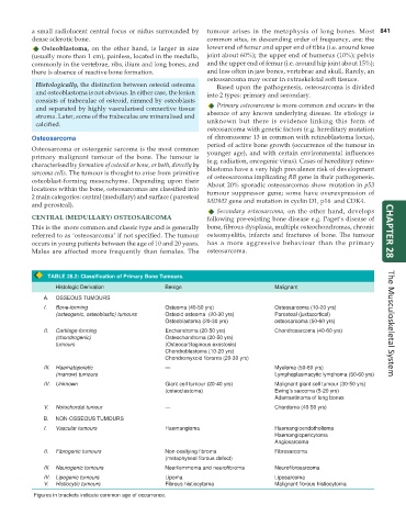
TABLE 28.2: Classification of Primary Bone Tumours.
Histologic Derivation Benign Malignant
A. OSSEOUS TUMOURS
I. Bone-forming Osteoma (40-50 yrs) Osteosarcoma (10-20 yrs)
(osteogenic, osteoblastic) tumours Osteoid osteoma (20-30 yrs) Parosteal (juxtacortical)
Osteoblastoma (20-30 yrs) osteosarcoma (50-60 yrs) The Musculoskeletal System
II. Cartilage-forming Enchondroma (20-50 yrs) Chondrosarcoma (40-60 yrs)
(chondrogenic) Osteochondroma (20-50 yrs)
tumours (Osteocartilaginous exostosis)
Chondroblastoma (10-20 yrs)
Chondromyxoid fibroma (20-30 yrs)
III. Haematopoietic — Myeloma (50-60 yrs)
(marrow) tumours Lymphoplasmacytic lymphoma (50-60 yrs)
IV. Unknown Giant cell tumour (20-40 yrs) Malignant giant cell tumour (30-50 yrs)
(osteoclastoma) Ewing’s sarcoma (5-20 yrs)
Adamantinoma of long bones
V. Notochordal tumour — Chordoma (40-50 yrs)
B. NON-OSSEOUS TUMOURS
I. Vascular tumours Haemangioma Haemangioendothelioma
Haemangiopericytoma
Angiosarcoma
II. Fibrogenic tumours Non-ossifying fibroma Fibrosarcoma
(metaphyseal fibrous defect)
III. Neurogenic tumours Neurilemmoma and neurofibroma Neurofibrosarcoma
IV. Lipogenic tumours Lipoma Liposarcoma
V. Histiocytic tumours Fibrous histiocytoma Malignant fibrous histiocytoma
Figures in brackets indicate common age of occurrence.