
Page 854 - Textbook of Pathology, 6th Edition
P. 854
838 of replacement of bone by fibrous connective tissue with a
characteristic whorled pattern and containing trabeculae of
woven bone. Radiologically, the typical focus of fibrous
dysplasia has well-demarcated ground-glass appearance.
Three types of fibrous dysplasia are distinguished—
monostotic, polyostotic, and Albright syndrome. The
spectrum of phenotype of the disease is due to activating
mutation in GNAS1 gene, which encodes for α-subunits of
the stimulatory G-protein, G .
Sα
Monostotic fibrous dysplasia. Monostotic fibrous
dysplasia affects a solitary bone and is the most common
type, comprising about 70% of all cases. The condition affects
either sex and most patients are between 20 and 30 years of
age. The bones most often affected, in descending order of
frequency, are: ribs, craniofacial bones (especially maxilla),
femur, tibia and humerus. The condition generally remains
asymptomatic and is discovered incidentally, but
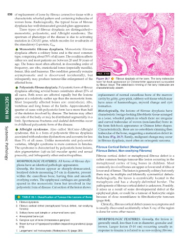
infrequently may produce tumour-like enlargement of the Figure 28.7 Fibrous dysplasia of the bone. The bony trabeculae
affected bone. have fish-hook appearance (or Chinese-letter appearance) surrounded
by fibrous tissue. The osteoblastic rimming of the bony trabeculae are
Polyostotic fibrous dysplasia. Polyostotic form of fibrous characteristically absent.
dysplasia affecting several bones constitutes about 25% of
all cases. Both sexes are affected equally but the lesions replacement of normal cancellous bone of the marrow
appear at a relatively earlier age than the monostotic form. cavity by gritty, grey-pink, rubbery soft tissue which may
Most frequently affected bones are: craniofacial, ribs, have areas of haemorrhages, myxoid change and cyst
vertebrae and long bones of the limbs. Approximately a formation.
quarter of cases with polyostotic form have more than half Histologically, the lesions of fibrous dysplasia have
of the skeleton involved by disease. The lesions may affect characteristic benign-looking fibroblastic tissue arranged
one side of the body or may be distributed segmentally in a in a loose, whorled pattern in which there are irregular
limb. Spontaneous fractures and skeletal deformities occur and curved trabeculae of woven (non-lamellar) bone in
SECTION III
in childhood polyostotic form of the disease. the form fish-hook appearance or Chinese letter shapes.
Albright syndrome. Also called McCune-Albright Characteristically, there are no osteoblasts rimming then
syndrome, this is a form of polyostotic fibrous dysplasia trabeculae of the bone, suggesting a maturation defect in
associated with endocrine dysfunctions and accounts for less the bone (Fig. 28.7). Rarely, malignant change may occur
than 5% of all cases. Unlike monostotic and polyostotic in fibrous dysplasia, most often an osteogenic sarcoma.
varieties, Albright syndrome is more common in females.
The syndrome is characterised by polyostotic bone lesions, Fibrous Cortical Defect (Metaphyseal
skin pigmentation (cafe-au-lait macular spots) and sexual Fibrous Defect, Non-ossifying Fibroma)
precocity, and infrequently other endocrinopathies. Fibrous cortical defect or metaphyseal fibrous defect is a
rather common benign tumour-like lesion occurring in the
MORPHOLOGIC FEATURES. All forms of fibrous dys- metaphyseal cortex of long bones in children. Most
Systemic Pathology
plasia have an identical pathologic appearance. commonly involved bones are upper or lower end of tibia or
Grossly, the lesions appear as sharply-demarcated, lower end of femur. The lesion is generally solitary but rarely
localised defects measuring 2-5 cm in diameter, present there may be multiple and bilaterally symmetrical defects.
within the cancellous bone, having thin and smooth Radiologically, the lesion is eccentrically located in the
overlying cortex. The epiphyseal cartilages are generally metaphysis and has a sharply-delimited border. The
spared in the monostotic form but involved in the pathogenesis of fibrous cortical defect is unknown. Possibly,
polyostotic form of disease. Cut section of the lesion shows
it arises as a result of some developmental defect at the
epiphyseal plate, or could be a tumour of histiocytic origin
TABLE 28.1: Classification of Tumour-like Lesions of Bone. because of close resemblance to fibrohistiocytic tumours
(page 864).
1. Fibrous dysplasia Clinically, fibrous cortical defect causes no symptoms and
2. Fibrous cortical defect (metaphyseal fibrous defect, non-ossifying is usually discovered accidentally when X-ray of the region
fibroma)
3. Solitary bone cyst (simple or unicameral bone cyst) is done for some other reason.
4. Aneurysmal bone cyst
5. Ganglion cyst of bone (intraosseous ganglion) MORPHOLOGIC FEATURES. Grossly, the lesion is
6. Brown tumour of hyperparathyroidism (reparative granuloma) (page generally small, less than 4 cm in diameter, granular and
816) brown. Larger lesion (5-10 cm) occurring usually in
7. Langerhans’ cell histiocytosis (Histiocytosis-X) (page 385) response to trauma is referred to as non-ossifying fibroma.