
Page 884 - Textbook of Pathology, 6th Edition
P. 884
868
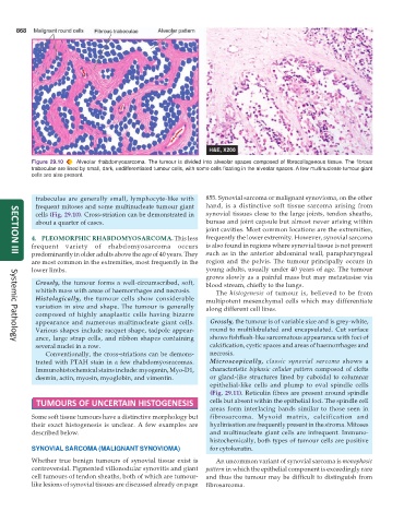
Figure 29.10 Alveolar rhabdomyosarcoma. The tumour is divided into alveolar spaces composed of fibrocollagenous tissue. The fibrous
trabeculae are lined by small, dark, undifferentiated tumour cells, with some cells floating in the alveolar spaces. A few multinucleate tumour giant
cells are also present.
trabeculae are generally small, lymphocyte-like with 855. Synovial sarcoma or malignant synovioma, on the other
frequent mitoses and some multinucleate tumour giant hand, is a distinctive soft tissue sarcoma arising from
cells (Fig. 29.10). Cross-striation can be demonstrated in synovial tissues close to the large joints, tendon sheaths,
about a quarter of cases. bursae and joint capsule but almost never arising within
joint cavities. Most common locations are the extremities,
4. PLEOMORPHIC RHABDOMYOSARCOMA. This less frequently the lower extremity. However, synovial sarcoma
frequent variety of rhabdomyosarcoma occurs is also found in regions where synovial tissue is not present
predominantly in older adults above the age of 40 years. They such as in the anterior abdominal wall, parapharyngeal
SECTION III
are most common in the extremities, most frequently in the region and the pelvis. The tumour principally occurs in
lower limbs. young adults, usually under 40 years of age. The tumour
grows slowly as a painful mass but may metastasise via
Grossly, the tumour forms a well-circumscribed, soft, blood stream, chiefly to the lungs.
whitish mass with areas of haemorrhages and necrosis. The histogenesis of tumour is, believed to be from
Histologically, the tumour cells show considerable multipotent mesenchymal cells which may differentiate
variation in size and shape. The tumour is generally along different cell lines.
composed of highly anaplastic cells having bizarre
appearance and numerous multinucleate giant cells. Grossly, the tumour is of variable size and is grey-white,
Various shapes include racquet shape, tadpole appear- round to multilobulated and encapsulated. Cut surface
ance, large strap cells, and ribbon shapes containing shows fishflesh-like sarcomatous appearance with foci of
Systemic Pathology
several nuclei in a row. calcification, cystic spaces and areas of haemorrhages and
Conventionally, the cross-striations can be demons- necrosis.
trated with PTAH stain in a few rhabdomyosracomas. Microscopically, classic synovial sarcoma shows a
Immunohistochemical stains include: myogenin, Myo-D1, characteristic biphasic cellular pattern composed of clefts
desmin, actin, myosin, myoglobin, and vimentin. or gland-like structures lined by cuboidal to columnar
epithelial-like cells and plump to oval spindle cells
(Fig. 29.11). Reticulin fibres are present around spindle
TUMOURS OF UNCERTAIN HISTOGENESIS cells but absent within the epithelial foci. The spindle cell
areas form interlacing bands similar to those seen in
Some soft tissue tumours have a distinctive morphology but fibrosarcoma. Myxoid matrix, calcification and
their exact histogenesis is unclear. A few examples are hyalinisation are frequently present in the stroma. Mitoses
described below. and multinucleate giant cells are infrequent. Immuno-
histochemically, both types of tumour cells are positive
SYNOVIAL SARCOMA (MALIGNANT SYNOVIOMA) for cytokeratin.
Whether true benign tumours of synovial tissue exist is An uncommon variant of synovial sarcoma is monophasic
controversial. Pigmented villonodular synovitis and giant pattern in which the epithelial component is exceedingly rare
cell tumours of tendon sheaths, both of which are tumour- and thus the tumour may be difficult to distinguish from
like lesions of synovial tissues are discussed already on page fibrosarcoma.