
Page 879 - Textbook of Pathology, 6th Edition
P. 879
blasts which have high mitotic rate. Ultrastructurally, 863
some of the fibroblasts have features of myofibroblasts
having contractile nature. The palmar lesions frequently
extend into soft tissues causing contractures. Both palmar
and plantar lesions may remain stationary at nodular
stage, progress, or regress spontaneously. Recurrence rate
after surgical excision in both forms is as high as 50-60%.
DESMOID FIBROMATOSES. Desmoid fibromatoses or
musculo-aponeurotic fibromatoses, commonly referred to as
desmoid tumours, are of 2 types: abdominal and extra-
abdominal. Both types are, however, histologically similar.
Clinically, both types behave in an aggressive manner and
have to be distinguished from sarcomas. Recurrences are
frequent and multiple. The pathogenesis of these lesions is
not known but among the factors implicated are the role of
antecedent trauma, genetic influences and relationship to
oestrogen as obsereved by occurrence of these lesions in Figure 29.2 Fibrosarcoma, common clinical location.
pregnancy.
Abdominal desmoids are locally aggressive infiltrating are composed of uniform-looking fibroblasts arranged in
tumour-like fibroblastic growths, often found in the musculo- bands and fascicles. Pleomorphism and mitoses are
aponeurotic structures of the rectus muscle in the anterior infrequent. The older regions of the tumour have hypo-
abdominal wall in women during or after pregnancy.
cellular hyalinised collagen.
Extra-abdominal desmoids, on the other hand, are more
common in men and are widely distributed such as in the CHAPTER 29
upper and lower extremities, chest wall, back, buttocks, and FIBROSARCOMA
head and neck region. The number of soft tissue tumours diagnosed as fibrosarcoma
Intra-abdominal desmoids present at the root of the small has now dropped, partly because of reclassification of
bowel mesentery are associated with Gardner’s syndrome fibromatoses which have aggressive and recurrent behaviour,
(consisting of fibromatosis, familial intestinal polyposis, and partly due to inclusion of many of such tumours in the
osteomas and epidermal cysts). group of fibrous histiocytomas (described later).
Fibrosarcoma is a slow-growing tumour, affecting adults
Grossly, desmoids are solitary, large, grey-white, firm and between 4th and 7th decades of life. Most common locations
unencapsulated tumours infiltrating the muscle locally. are the lower extremity (especially thigh and around the
Cut surface is whorled and trabeculated. knee), upper extremity, trunk, head and neck, and Soft Tissue Tumours
Microscopically, their appearance is rather misleadingly retroperitoneum (Fig. 29.2). The tumour is capable of
bland in contrast with aggressive local behaviour. They metastasis, chiefly via the blood stream.
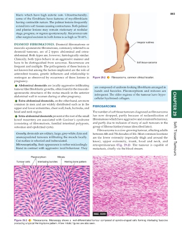
Figure 29.3 Fibrosarcoma. Microscopy shows a well-differentiated tumour composed of spindle-shaped cells forming interlacing fascicles
producing a typical Herring-bone pattern. A few mitotic figures are also seen.