
Page 880 - Textbook of Pathology, 6th Edition
P. 880
864 Grossly, fibrosarcoma is a grey-white, firm, lobulated and
characteristically circumscribed mass. Cut surface of the
tumour is soft, fishflesh-like, with foci of necrosis and
haemorrhages.
Histologically, the tumour is composed of uniform,
spindle-shaped fibroblasts arranged in intersecting
fascicles. In well-differentiated tumours, such areas
produce ‘herring-bone pattern’ (herring-bone is a sea fish)
(Fig. 29.3). Poorly-differentiated fibrosarcoma, however,
has highly pleomorphic appearance with frequent mitoses
and bizarre cells.
FIBROHISTIOCYTIC TUMOURS
The group of fibrohistiocytic tumours is characterised by
distinctive light microscopic features that include presence
of cells with fibroblastic and histiocytic features in varying
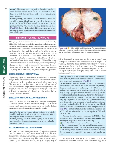
proportion and identification of characteristic cart-wheel or Figure 29.4 Malignant fibrous histiocytoma. The lobulated tumour
storiform pattern in which the spindle cells radiate outward infiltrating the skeletal muscle is somewhat circumscribed. Cut surface
is grey-white fleshy with areas of haemorrhage and necrosis.
from the central focus. The histogenesis of these cells is
uncertain but possibly they arise from primitive
mesenchymal cells or facultative fibroblasts which are
capable of differentiating along different cell lines. The group 5th to 7th decades. Most common locations are the lower
includes full spectrum of lesions varying from benign (benign and upper extremities and retroperitoneum. It begins as a
painless, enlarging mass, generally in relation to skeletal
fibrous histiocytoma) to malignant (malignant fibrous muscle, deep fascia or subcutaneous tissue. The tumour is
histiocytoma), with dermatofibrosarcoma protuberans believed to arise from primitive mesenchymal cells which
occupying the intermediate (low-grade malignancy) position.
are capable of differentiating towards both fibroblastic and
histiocytic cell lines.
SECTION III
BENIGN FIBROUS HISTIOCYTOMA
Grossly, MFH is a multilobulated, well-circumscribed,
Depending upon the location and predominant pattern, firm or fleshy mass, 5-10 cm in diameter. Cut surface is
benign fibrous histiocytomas include a number of diverse grey-white, soft and myxoid (Fig. 29.4).
entities such as dermatofibroma, sclerosing haemangioma, Histologically, there is marked variation in appearance
fibroxanthoma, xanthogranuloma, giant cell tumour of
tendon sheath and pigmented villonodular synovitis. All from area to area within the same tumour. In general,
these tumours have mixed composition of benign fibroblastic there is admixture of spindle-shaped fibroblast-like cells
and histiocytic pattern of cells and have been described in and mononuclear round to oval histiocyte-like cells which
relevant sections already. may show phagocytic function. There is tendency for the
spindle shaped cells to be arranged in characteristic cart-
DERMATOFIBROSARCOMA PROTUBERANS wheel or storiform pattern. The tumour cells show
Systemic Pathology
varying degree of pleomorphism, hyperchromatism,
Dermatofibrosarcoma protuberans is a low-grade malignant mitotic activity and presence of multinucleate bizarre
cutaneous tumour of fibrohistiocytic origin. The tumour tumour giant cells. Usually there are numerous blood
recurs locally, and in rare instances gives rise to distant vessels and some scattered lymphocytes and plasma cells
metastases. Most frequent location is the trunk. (Fig. 29.5). Important immunohistochemical markers for
Grossly, the tumour forms a firm, solitary or multiple, MFH include vimentin, α-chymotrypsin, CD68 and
satellite nodules extending into the subcutaneous fat and factor VIII-a.
having thin and ulcerated skin surface. Besides the storifrom pleomorphic MFH as the
Histologically, the tumour is highly cellular and is prototype, a few morphologic variants of MFH having
composed of fibroblasts arranged in a cart-wheel or bearing on prognosis include the following:
storiform pattern. Myxoid MFH shows areas of loose myxoid stroma
in the cellular areas and has an overall better prognosis.
MALIGNANT FIBROUS HISTIOCYTOMA Inflammatory MFH is an undifferentiated high-grade
MFH having prominent neutrophilic infiltrate besides
Malignant fibrous histiocytomas (MFH) represent approxi- the presence of eosinophils, histiocytes and xanthoma
mately 20-30% of all soft tissue sarcomas. It is the most cells.
common soft tissue sarcoma and is the most frequent sarcoma
associated with radiotherapy. The tumour occurs more Prognosis is determined by 2 parameters: depth of
commonly in males and more frequently in the age group of location and size of the tumour. Deep-seated and large MFH