
Page 882 - Textbook of Pathology, 6th Edition
P. 882
866
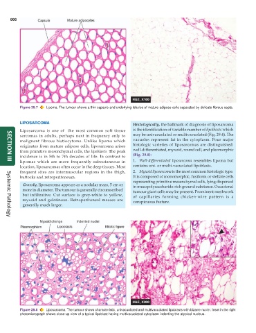
Figure 29.7 Lipoma. The tumour shows a thin capsule and underlying lobules of mature adipose cells separated by delicate fibrous septa.
LIPOSARCOMA Histologically, the hallmark of diagnosis of liposarcoma
Liposarcoma is one of the most common soft tissue is the identification of variable number of lipoblasts which
sarcomas in adults, perhaps next in frequency only to may be univacuolated or multivacuolated (Fig. 29.4). The
malignant fibrous histiocytoma. Unlike lipoma which vacuoles represent fat in the cytoplasm. Four major
originates from mature adipose cells, liposarcoma arises histologic varieties of liposarcomas are distinguished:
from primitive mesenchymal cells, the lipoblasts. The peak well-differentiated, myxoid, round cell, and pleomorphic
incidence is in 5th to 7th decades of life. In contrast to (Fig. 29.8):
lipomas which are more frequently subcutaneous in 1. Well-differentiated liposarcoma resembles lipoma but
SECTION III
location, liposarcomas often occur in the deep tissues. Most contains uni- or multi-vacuolated lipoblasts.
frequent sites are intermuscular regions in the thigh, 2. Myxoid liposarcoma is the most common histologic type.
buttocks and retroperitoneum. It is composed of monomorphic, fusiform or stellate cells
representing primitive mesenchymal cells, lying dispersed
Grossly, liposarcoma appears as a nodular mass, 5 cm or in mucopolysaccharide-rich ground substance. Occasional
more in diameter. The tumour is generally circumscribed tumour giant cells may be present. Prominent meshwork
but infiltrative. Cut surface is grey-white to yellow, of capillaries forming chicken-wire pattern is a
myxoid and gelatinous. Retroperitoneal masses are conspicuous feature.
generally much larger.
Systemic Pathology
Figure 29.8 Liposarcoma. The tumour shows characteristic, univacuolated and multivacuolated lipoblasts with bizarre nuclei. Inset in the right
photomicrograph shows close-up view of a typical lipoblast having multivacuolated cytoplasm indenting the atypical nucleus.