
Page 890 - Textbook of Pathology, 6th Edition
P. 890
874 SECONDARY HYDROCEPHALUS. Secondary hydro-
cephalus is much less common and is defined as compen-
satory increase of CSF due to loss of neural tissue without
associated rise in intracranial pressure (normal pressure
hydrocephalus) e.g. from cerebral atrophy and infarction.
MORPHOLOGIC FEATURES. Grossly, there is dilatation
of the ventricles depending upon the site of obstruction.
There is thinning and stretching of the brain. The scalp
veins overlying the enlarged head are engorged and the
fontanelle remain open.
Histologically, severe hydrocephalus may be associated
with damage to ependymal lining of the ventricles and
periventricular interstitial oedema.
INFECTIONS
A large number of pathogens comprising various kinds of
bacteria, fungi, viruses, rickettsiae and parasites can cause
infections of the nervous system. The micro-organisms may
gain entry into the nervous system by one of the following
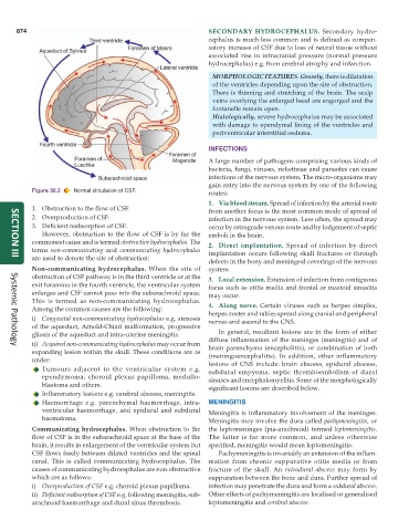
Figure 30.2 Normal circulation of CSF. routes:
1. Via blood stream. Spread of infection by the arterial route
1. Obstruction to the flow of CSF. from another focus is the most common mode of spread of
2. Overproduction of CSF. infection in the nervous system. Less often, the spread may
3. Deficient reabsorption of CSF. occur by retrograde venous route and by lodgement of septic
However, obstruction to the flow of CSF is by far the emboli in the brain.
commonest cause and is termed obstructive hydrocephalus. The 2. Direct implantation. Spread of infection by direct
terms non-communicating and communicating hydrocephalus implantation occurs following skull fractures or through
are used to denote the site of obstruction:
defects in the bony and meningeal coverings of the nervous
SECTION III
Non-communicating hydrocephalus. When the site of system.
obstruction of CSF pathway is in the third ventricle or at the 3. Local extension. Extension of infection from contiguous
exit foramina in the fourth ventricle, the ventricular system focus such as otitis media and frontal or mastoid sinusitis
enlarges and CSF cannot pass into the subarachnoid space. may occur.
This is termed as non-communicating hydrocephalus.
Among the common causes are the following: 4. Along nerve. Certain viruses such as herpes simplex,
herpes zoster and rabies spread along cranial and peripheral
i) Congenital non-communicating hydrocephalus e.g. stenosis nerves and ascend to the CNS.
of the aqueduct, Arnold-Chiari malformation, progressive
gliosis of the aqueduct and intra-uterine meningitis. In general, resultant lesions are in the form of either
ii) Acquired non-communicating hydrocephalus may occur from diffuse inflammation of the meninges (meningitis) and of
brain parenchyma (encephalitis), or combination of both
Systemic Pathology
expanding lesion within the skull. These conditions are as (meningoencephalitis). In addition, other inflammatory
under: lesions of CNS include: brain abscess, epidural abscess,
Tumours adjacent to the ventricular system e.g. subdural empyema, septic thromboembolism of dural
ependymoma, choroid plexus papilloma, medullo- sinuses and encephalomyelitis. Some of the morphologically
blastoma and others. significant lesions are described below.
Inflammatory lesions e.g. cerebral abscess, meningitis.
Haemorrhage e.g. parenchymal haemorrhage, intra- MENINGITIS
ventricular haemorrhage, and epidural and subdural Meningitis is inflammatory involvement of the meninges.
haematoma. Meningitis may involve the dura called pachymeningitis, or
Communicating hydrocephalus. When obstruction to the the leptomeninges (pia-arachnoid) termed leptomeningitis.
flow of CSF is in the subarachnoid space at the base of the The latter is far more common, and unless otherwise
brain, it results in enlargement of the ventricular system but specified, meningitis would mean leptomeningitis.
CSF flows freely between dilated ventricles and the spinal Pachymeningitis is invariably an extension of the inflam-
canal. This is called communicating hydrocephalus. The mation from chronic suppurative otitis media or from
causes of communicating hydrocephalus are non-obstructive fracture of the skull. An extradural abscess may form by
which are as follows: suppuration between the bone and dura. Further spread of
i) Overproduction of CSF e.g. choroid plexus papilloma. infection may penetrate the dura and form a subdural abscess.
ii) Deficient reabsorption of CSF e.g. following meningitis, sub- Other effects of pachymeningitis are localised or generalised
arachnoid haemorrhage and dural sinus thrombosis. leptomeningitis and cerebral abscess.