
Page 892 - Textbook of Pathology, 6th Edition
P. 892
876 4. CSF protein usually normal or mildly raised.
5. CSF sugar concentration usually normal.
6. CSF bacteriologically sterile.
Chronic (Tuberculous and Cryptococcal) Meningitis
There are two principal types of chronic meningitis—one
bacterial (tuberculous meningitis) and the other fungal
(cryptococcal meningitis). Both types cause chronic
granulomatous reaction and may produce parenchymal
lesions.
Tuberculous meningitis occurs in children and adults
through haematogenous spread of infection from tuber-
culosis elsewhere in the body, or it may simply be a mani-
festation of miliary tuberculosis. Less commonly, the spread
may occur directly from tuberculosis of a vertebral body.
Cryptococcal meningitis develops particularly in debilitated
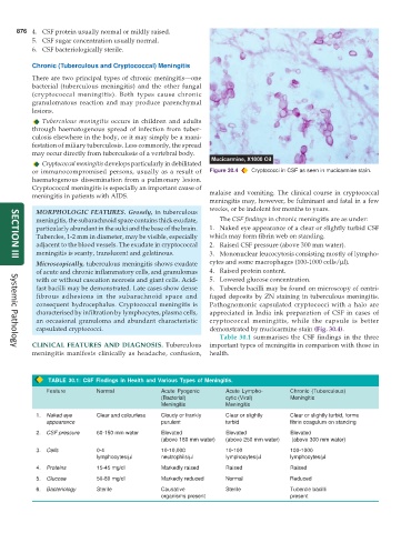
or immunocompromised persons, usually as a result of Figure 30.4 Cryptococci in CSF as seen in mucicarmine stain.
haematogenous dissemination from a pulmonary lesion.
Cryptococcal meningitis is especially an important cause of
meningitis in patients with AIDS. malaise and vomiting. The clinical course in cryptococcal
meningitis may, however, be fulminant and fatal in a few
weeks, or be indolent for months to years.
MORPHOLOGIC FEATURES. Grossly, in tuberculous
meningitis, the subarachnoid space contains thick exudate, The CSF findings in chronic meningitis are as under:
particularly abundant in the sulci and the base of the brain. 1. Naked eye appearance of a clear or slightly turbid CSF
Tubercles, 1-2 mm in diameter, may be visible, especially which may form fibrin web on standing.
adjacent to the blood vessels. The exudate in cryptococcal 2. Raised CSF pressure (above 300 mm water).
meningitis is scanty, translucent and gelatinous. 3. Mononuclear leucocytosis consisting mostly of lympho-
SECTION III
Microscopically, tuberculous meningitis shows exudate cytes and some macrophages (100-1000 cells/μl).
of acute and chronic inflammatory cells, and granulomas 4. Raised protein content.
with or without caseation necrosis and giant cells. Acid- 5. Lowered glucose concentration.
fast bacilli may be demonstrated. Late cases show dense 6. Tubercle bacilli may be found on microscopy of centri-
fibrous adhesions in the subarachnoid space and fuged deposits by ZN staining in tuberculous meningitis.
consequent hydrocephalus. Cryptococcal meningitis is Pathognomonic capsulated cryptococci with a halo are
characterised by infiltration by lymphocytes, plasma cells, appreciated in India ink preparation of CSF in cases of
an occasional granuloma and abundant characteristic cryptococcal meningitis, while the capsule is better
capsulated cryptococci. demonstrated by mucicarmine stain (Fig. 30.4).
Table 30.1 summarises the CSF findings in the three
CLINICAL FEATURES AND DIAGNOSIS. Tuberculous important types of meningitis in comparison with those in
Systemic Pathology
meningitis manifests clinically as headache, confusion, health.
TABLE 30.1: CSF Findings in Health and Various Types of Meningitis.
Feature Normal Acute Pyogenic Acute Lympho- Chronic (Tuberculous)
(Bacterial) cytic (Viral) Meningitis
Meningitis Meningitis
1. Naked eye Clear and colourless Cloudy or frankly Clear or slightly Clear or slightly turbid, forms
appearance purulent turbid fibrin coagulum on standing
2. CSF pressure 60-150 mm water Elevated Elevated Elevated
(above 180 mm water) (above 250 mm water) (above 300 mm water)
3. Cells 0-4 10-10,000 10-100 100-1000
lymphocytes/μl neutrophils/μl lymphocytes/μl lymphocytes/μl
4. Proteins 15-45 mg/dl Markedly raised Raised Raised
5. Glucose 50-80 mg/dl Markedly reduced Normal Reduced
6. Bacteriology Sterile Causative Sterile Tubercle bacilli
organisms present present