
Page 893 - Textbook of Pathology, 6th Edition
P. 893
ENCEPHALITIS 877
Parenchymal infection of brain is termed encephalitis.
Encephalitis may be the result of bacterial, viral, fungal and
protozoal infections.
Bacterial Encephalitis
Bacterial infection of the brain substance is usually secondary
to involvement of the meninges rather than a primary
bacterial parenchymal infection. This results in bacterial
cerebritis that progresses to form brain abscess. However,
tuberculosis and neurosyphilis are the two primary bacterial
involvements of the brain parenchyma.
BRAIN ABSCESS. Brain abscesses may arise by one of the
following routes:
1. By direct implantation of organisms e.g. following
compound fractures of the skull.
2. By local extension of infection e.g. chronic suppurative
otitis media, mastoiditis and sinusitis.
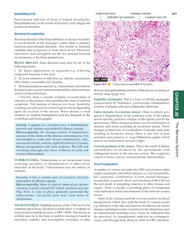
Figure 30.5 Tuberculous encephalitis of the brain.
3. By haematogenous spread e.g. from primary infection in
the heart such as acute bacterial endocarditis, and from lungs dorsalis and generalised paralysis of the insane occurring in
such as in bronchiectasis. tertiary stage (page 161).
Clinically, there is usually evidence of reactivation of
infection at the primary site preceding the onset of cerebral Syphilitic meningitis. This is a form of chronic meningitis
symptoms. The features of abscess are fever, headache, characterised by distinctive perivascular inflammatory CHAPTER 30
vomiting, seizures and focal neurological deficits depending reaction of plasma cells and endarteritis obliterans.
upon the location of the abscess. Brain abscess is most Tabes dorsalis (Locomotor ataxia). There is slowly pro-
common in cerebral hemispheres and less frequent in the gressive degeneration of the posterior roots of the spinal
cerebellum and basal ganglia. nerves and the posterior columns of the spinal cord by the
spirochaetes. These changes produce loss of coordination of
Grossly, it appears as a localised area of inflammatory muscles and joints resulting in locomotor ataxia. These
necrosis and oedema surrounded by fibrous capsule. changes produce loss of coordination of muscles and joints
Microscopically, the changes consist of liquefactive resulting in locomotor ataxia. There is also loss of pain
necrosis in the centre of the abscess containing pus. It is sensation and presence of Argyll-Robertson pupils which
surrounded by acute and chronic inflammatory cells, react to accommodation but not to light. The Nervous System
neovascularisation, oedema, septic thrombosis of vessels,
fibrous encapsulation and zone of gliosis. The CSF and General paralysis of the insane. This is the result of diffuse
overlying meninges also show evidence of acute and parenchymal involvement by the spirochaetes with
chronic inflammation. widespread lesions in the nervous system. The symptoms
consist of motor, sensory and psychiatric abnormalities.
TUBERCULOMA. Tuberculoma is an intracranial mass
occurring secondary to dissemination of tuberculosis Viral Encephalitis
elsewhere in the body. Tuberculomas may be solitary or
multiple. A number of viruses can infect the CNS and produce either
aseptic meningitis (described already) or viral encephalitis,
Grossly, it has a central area of caseation necrosis but sometimes combination of both termed meningo-
surrounded by fibrous capsule. encephalitis, is present. Most viral infections of the CNS are
Microscopically, there is typical tuberculous granu- the end-result of preceding infection in other tissues and
lomatous reaction around the central caseation necrosis organs. There is usually a preceding phase of extraneural
(Fig. 30.5). A zone of gliosis generally surrounds the viral replication before involvement of the nervous system
tuberculoma. Advanced cases may show areas of occurs.
calcification. Most of the viruses reach the nervous system via blood
stream before which they enter the body by various routes
NEUROSYPHILIS. Syphilitic lesions of the CNS used to be e.g. infection of the skin and mucous membrane (in herpes
common and serious, but more recently there is evidence of simplex and herpes zoster-varicella), by the alimentary tract
atypical neurosyphilis in cases of HIV/AIDS. The lesions in (in enteroviruses including polio virus), by arthropod bite
syphilis may be in the form of syphilitic meningitis found in (in arbovirus), by transplacental infection (in cytomegalo-
secondary syphilis, and neurosyphilis consisting of tabes virus), and through body fluids in AIDS (in HIV infection).