
Page 897 - Textbook of Pathology, 6th Edition
P. 897
881
Figure 30.7 An old cystic infarct in the brain (coronal section). There
is shrinkage of scarred area with ipsilateral ventricular dilatation.
becomes evident 6-12 hours after its occurrence. The
affected area is soft and swollen and there is blurring of
junction between grey and white matter. Within 2-3 days,
the infarct undergoes softening and disintegration.
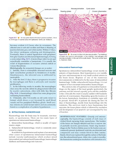
Eventually, there is central liquefaction with peripheral Figure 30.8 An anaemic infarct of a few days duration. The histologic
firm glial reaction and thickened leptomeninges, forming changes are reactive astrocytosis, a few reactive macrophages and
a cystic infarct (Fig. 30.7). A haemorrhagic infarct is red and neovascularisation in the wall of the cystic lesion. The outer cortical layer
superficially resembles a haematoma. It is usually the is, however, intact.
result of fragmentation of occlusive arterial emboli or
venous thrombosis. CHAPTER 30
Histologically, the sequential changes are as under: Intracerebral Haemorrhage
1. Initially, there is eosinophilic neuronal necrosis and Spontaneous intracerebral haemorrhage occurs mostly in
lipid vacuolisation produced by breakdown of myelin. patients of hypertension. Most hypertensives over middle
Simultaneously, the infarcted area is infiltrated by age have microaneurysms in very small cerebral arteries in
neutrophils. the brain tissue. Rupture of one of the numerous
2. After the first 2-3 days, there is progressive invasion microaneurysms is believed to be the cause of intracerebral
by macrophages and there is astrocytic and vascular haemorrhage. Unlike subarachnoid haemorrhage, it is not
proliferation. common to have recurrent intracerebral haemorrhages.
3. In the following weeks to months, the macrophages The common sites of hypertensive intracerebral haemor-
clear away the necrotic debris by phagocytosis followed rhage are the region of the basal ganglia (particularly the The Nervous System
by reactive astrocytosis, often with little fine fibrosis putamen and the internal capsule), pons and the cerebellar
(Fig. 30.8). A haemorrhagic infarct has some phagocytes cortex. Clinically the onset is usually sudden with headache
containing haemosiderin. and loss of consciousness. Depending upon the location of
4. Ultimately, after 3-4 months an old cystic infarct is the lesion, hemispheric, brainstem or cerebellar signs will
formed which shows a cyst traversed by small blood be present. About 40% of patients die during the first 3-4
vessels and has peripheral fibrillary gliosis. Small cavi- days of haemorrhage, mostly from haemorrhage into the
tary infarcts are called lacunar infarcts and are commonly ventricles. The survivors tend to have haematoma that
found as a complication of systemic hypertension. separates the tissue planes which is followed by resolution
and development of an apoplectic cyst accompanied by loss
B. INTRACRANIAL HAEMORRHAGE of function.
Haemorrhage into the brain may be traumatic, non-trau- MORPHOLOGIC FEATURES. Grossly and micros-
matic, or spontaneous. There are two main types of copically, the haemorrhage consists of dark mass of
spontaneous intracranial haemorrhage: clotted blood replacing brain parenchyma. The borders
1. Intracerebral haemorrhage, which is usually of hyper- of the lesion are sharply-defined and have a narrow rim
tensive origin. of partially necrotic parenchyma. Small ring haemor-
2. Subarachnoid haemorrhage, which is commonly aneu- rhages in the Virchow-Robin space in the border zone are
rysmal in origin. commonly present. Ipsilateral ventricles are distorted and
In addition to hypertension and rupture of an aneurysm, compressed and may contain blood in their lumina.
other causes of spontaneous intracranial haemorrhage Rarely, blood may rupture through the surface of the brain
include vascular malformations which produce mixed into the subarachnoid space. After a few weeks to months,
intracerebral and subarachnoid haemorrhage, haemorrhagic the haematoma undergoes resolution with formation of a
diathesis and haemorrhage into tumours.