
Page 898 - Textbook of Pathology, 6th Edition
P. 898
882 slit-like space called apoplectic cyst which contains The remaining 15% cases of subarachnoid haemorrhage
yellowish fluid. Its margins are yellow-brown and have are the result of rupture in the posterior circulation, vascular
haemosiderin-laden macrophages and a reactive zone of malformations and rupture of mycotic aneurysms that occurs
fibrillary astrocytosis. in the setting of bacterial endocarditis. In all types of
aneurysms, the rupture of thin-walled dilatation occurs in
Subarachnoid Haemorrhage association with sudden rise in intravascular pressure but
Haemorrhage into the subarachnoid space is most comm- chronic hypertension does not appear to be a risk factor in
only caused by rupture of an aneurysm, and rarely, rupture their development or rupture.
of a vascular malformation. Clinically, berry aneurysms remain asymptomatic prior
A general discussion of aneurysms is given on page 405. to rupture. On rupture, they produce severe generalised
Of the three types of aneurysms affecting the larger headache of sudden onset which is frequently followed by
unconsciousness and neurologic defects. Initial mortality
intracranial arteries—berry, mycotic and fusiform, berry from first rupture is about 20-25%. Survivors recover
aneurysms are most important and most common.
completely but frequently suffer from recurrent episodes of
BERRY ANEURYSMS are saccular in appearance with fresh bleeding.
rounded or lobulated bulge arising at the bifurcation of
intracranial arteries and varying in size from 2 mm to 2 cm MORPHOLOGIC FEATURES. Rupture of a berry aneu-
or more. They account for 95% of aneurysms which are liable rysm frequently spreads haemorrhage throughout the
to rupture. Berry aneurysms are rare in childhood but subarachnoid space with rise in intracranial pressure and
increase in frequency in young adults and middle life. They characteristic blood-stained CSF. An intracerebral
are, therefore, not congenital anomalies but develop over the haematoma may develop if the blood tracks into the brain
years from developmental defect of the media of the arterial parenchyma. The region of the brain supplied by the
wall at the bifurcation of arteries forming thin-walled saccu- affected artery frequently shows infarction, partly
lar bulges. Although most berry aneurysms are sporadic in attributed to vasospasm.
occurrence, there is an increased incidence of their presence
in association with congenital polycystic kidney disease and TRAUMA TO THE CNS
coarctation of the aorta. About a quarter of berry aneurysms
are multiple. Trauma to the CNS constitutes an important cause of death
In more than 85% cases of subarachnoid haemorrhage, and permanent disability in the modern world. Important
the cause is massive and sudden bleeding from a berry causes of head injuries are: motor vehicle accidents,
SECTION III
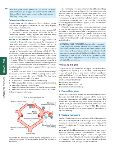
aneurysm on or near the circle of Willis. The four most accidental falls and violence. Traumatic injuries to the CNS
common sites are as under (Fig. 30.9): may result in three consequences which may occur in
1. In relation to anterior communicating artery. isolation or in combination:
2. At the origin of the posterior communicating artery from epidural haematoma;
the stem of the internal carotid artery. subdural haematoma; and
3. At the first major bifurcation of the middle cerebral artery. parenchymal brain damage.
4. At the bifurcation of the internal carotid into the middle
and anterior cerebral arteries. A. Epidural Haematoma
Epidural haematoma is accumulation of blood between the
dura and the skull following fracture of the skull, most
Systemic Pathology
commonly from rupture of middle meningeal artery. The
haematoma expands rapidly since accumulating blood is
arterial in origin and causes compression of the dura and
flattening of underlying gyri (Fig. 30.10). The patient
develops progressive loss of consciousness if haematoma is
not drained early.
B. Subdural Haematoma
Subdural haematoma is accumulation of blood between the
dura and subarachnoid and develops most often from
rupture of veins which cross the surface convexities of the
cerebral hemispheres. Subdural haematoma may be acute
or chronic.
Acute subdural haematoma. Acute subdural haematoma
develops following trauma and consists of clotted blood,
often in the frontoparietal region. There is no significant
compression of gyri (Fig. 30.11). Since the accumulated blood
Figure 30.9 The circle of Willis showing principal sites of berry
(saccular) aneurysms. The serial numbers indicate the frequency of is of venous origin, symptoms appear slowly and may
involvement. become chronic with passage of time if not fatal.