
Page 899 - Textbook of Pathology, 6th Edition
P. 899
883
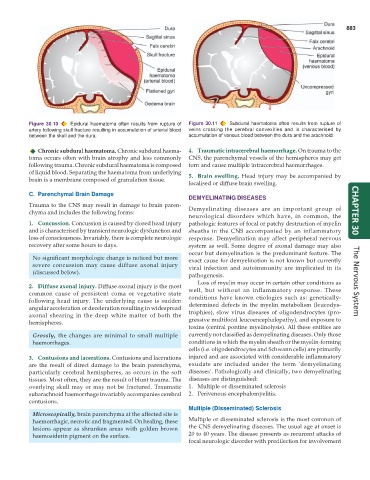
Figure 30.10 Epidural haematoma often results from rupture of Figure 30.11 Subdural haematoma often results from rupture of
artery following skull fracture resulting in accumulation of arterial blood veins crossing the cerebral convexities and is characterised by
between the skull and the dura. accumulation of venous blood between the dura and the arachnoid.
Chronic subdural haematoma. Chronic subdural haema- 4. Traumatic intracerebral haemorrhage. On trauma to the
toma occurs often with brain atrophy and less commonly CNS, the parenchymal vessels of the hemispheres may get
following trauma. Chronic subdural haematoma is composed torn and cause multiple intracerebral haemorrhages.
of liquid blood. Separating the haematoma from underlying 5. Brain swelling. Head injury may be accompanied by
brain is a membrane composed of granulation tissue.
localised or diffuse brain swelling.
C. Parenchymal Brain Damage
DEMYELINATING DISEASES
Trauma to the CNS may result in damage to brain paren- Demyelinating diseases are an important group of
chyma and includes the following forms: CHAPTER 30
neurological disorders which have, in common, the
1. Concussion. Concussion is caused by closed head injury pathologic features of focal or patchy destruction of myelin
and is characterised by transient neurologic dysfunction and sheaths in the CNS accompanied by an inflammatory
loss of consciousness. Invariably, there is complete neurologic response. Demyelination may affect peripheral nervous
recovery after some hours to days. system as well. Some degree of axonal damage may also
occur but demyelination is the predominant feature. The
No significant morphologic change is noticed but more exact cause for demyelination is not known but currently
severe concussion may cause diffuse axonal injury viral infection and autoimmunity are implicated in its
(discussed below).
pathogenesis.
Loss of myelin may occur in certain other conditions as The Nervous System
2. Diffuse axonal injury. Diffuse axonal injury is the most
common cause of persistent coma or vegetative state well, but without an inflammatory response. These
following head injury. The underlying cause is sudden conditions have known etiologies such as: genetically-
angular acceleration or deceleration resulting in widespread determined defects in the myelin metabolism (leucodys-
axonal shearing in the deep white matter of both the trophies), slow virus diseases of oligodendrocytes (pro-
hemispheres. gressive multifocal leucoencephalopathy), and exposure to
toxins (central pontine myelinolysis). All these entities are
Grossly, the changes are minimal to small multiple currently not classified as demyelinating diseases. Only those
haemorrhages. conditions in which the myelin sheath or the myelin-forming
cells (i.e. oligodendrocytes and Schwann cells) are primarily
3. Contusions and lacerations. Contusions and lacerations injured and are associated with considerable inflammatory
are the result of direct damage to the brain parenchyma, exudate are included under the term ‘demyelinating
particularly cerebral hemispheres, as occurs in the soft diseases’. Pathologically and clinically, two demyelinating
tissues. Most often, they are the result of blunt trauma. The diseases are distinguished:
overlying skull may or may not be fractured. Traumatic 1. Multiple or disseminated sclerosis
subarachnoid haemorrhage invariably accompanies cerebral 2. Perivenous encephalomyelitis.
contusions.
Multiple (Disseminated) Sclerosis
Microscopically, brain parenchyma at the affected site is
haemorrhagic, necrotic and fragmented. On healing, these Multiple or disseminated sclerosis is the most common of
lesions appear as shrunken areas with golden brown the CNS demyelinating diseases. The usual age at onset is
haemosiderin pigment on the surface. 20 to 40 years. The disease presents as recurrent attacks of
focal neurologic disorder with predilection for involvement