
Page 903 - Textbook of Pathology, 6th Edition
P. 903
887
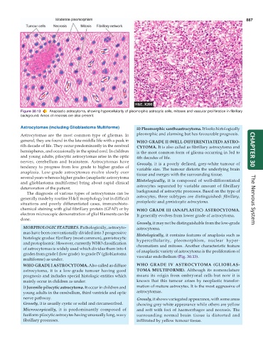
Figure 30.13 Anaplastic astrocytoma, showing hypercellularity of pleomorphic astrocytic cells, mitoses and vascular proliferation in fibrillary
background. Areas of necrosis are also present.
Astrocytomas (including Glioblastoma Multiforme) ii) Pleomorphic xanthoastrocytoma. It looks histologically
Astrocytomas are the most common type of gliomas. In pleomrphic and alarming but has favourable prognosis.
general, they are found in the late middle life with a peak in WHO GRADE II (WELL-DIFFERENTIATED) ASTRO-
6th decade of life. They occur predominantly in the cerebral CYTOMA. It is also called as fibrillary astrocytoma and
hemispheres, and occasionally in the spinal cord. In children is the most common form of glioma occurring in 3rd to CHAPTER 30
and young adults, pilocytic astrocytomas arise in the optic 4th decades of life.
nerves, cerebellum and brainstem. Astrocytomas have Grossly, it is a poorly defined, grey-white tumour of
tendency to progress from low grade to higher grades of variable size. The tumour distorts the underlying brain
anaplasia. Low-grade astrocytomas evolve slowly over tissue and merges with the surrounding tissue.
several years whereas higher grades (anaplastic astrocytoma
and glioblastoma multiforme) bring about rapid clinical Histologically, it is composed of well-differentiated
deterioration of the patient. astrocytes separated by variable amount of fibrillary
The diagnosis of various types of astrocytomas can be background of astrocytic processes. Based on the type of
generally made by routine H & E morphology but in difficult astrocytes, three subtypes are distinguished: fibrillary, The Nervous System
situations and poorly differentiated cases, immunohisto- protoplastic and gemistocytic astrocytoma.
chemical staining with glial fibrillary protein (GFAP) or by WHO GRADE III (ANAPLASTIC) ASTROCYTOMA.
electron microscopic demonstration of glial filaments can be It generally evolves from lower grade of astrocytoma.
done.
Grossly, it may not be distinguishable from the low-grade
MORPHOLOGIC FEATURES. Pathologically, astrocyto- astrocytoma.
mas have been conventionally divided into 3 progressive Histologically, it contains features of anaplasia such as
histologic grades: fibrillary (most common), gemistocytic hypercellularity, pleomorphism, nuclear hyper-
and protoplasmic. However, currently WHO classification chromatism and mitoses. Another characteristic feature
of astrocytomas is widely used which divides them into 4 of anaplastic variety of astrocytoma is the proliferation of
grades from grade I (low grade) to grade IV (glioblastoma vascular endothelium (Fig. 30.13).
multiforme) as under.
WHO GRADE I ASTROCYTOMA. Also called as diffuse WHO GRADE IV ASTROCYTOMA (GLIOBLAS-
astrocytoma, it is a low-grade tumour having good TOMA MULTIFORME). Although its nomenclature
prognosis and includes special histologic entities which means its origin from embryonal cells but now it is
mainly occur in children as under: known that this tumour arises by neoplastic transfor-
i) Juvenile pilocytic astrocytoma. It occur in children and mation of mature astrocytes. It is the most aggressive of
young adults in the cerebellum, third ventricle and optic astrocytomas.
nerve pathway. Grossly, it shows variegated appearance, with some areas
Grossly, it is usually cystic or solid and circumscribed. showing grey-white appearance while others are yellow
Microscopically, it is predominantly composed of and soft with foci of haemorrhages and necrosis. The
fusiform pilocytic astrocytes having unusually long, wavy surrounding normal brain tissue is distorted and
fibrillary processes. infiltrated by yellow tumour tissue.