
Page 904 - Textbook of Pathology, 6th Edition
P. 904
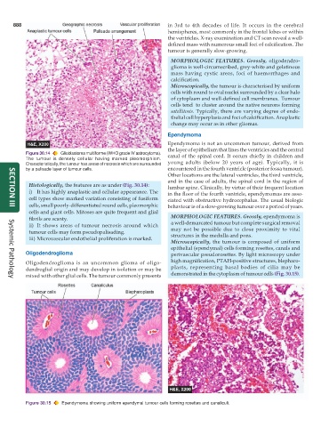
888                                                      in 3rd to 4th decades of life. It occurs in the cerebral
                                                               hemispheres, most commonly in the frontal lobes or within
                                                               the ventricles. X-ray examination and CT scan reveal a well-
                                                               defined mass with numerous small foci of calcification. The
                                                               tumour is generally slow-growing.
                                                                 MORPHOLOGIC FEATURES. Grossly, oligodendro-
                                                                 glioma is well-circumscribed, grey-white and gelatinous
                                                                 mass having cystic areas, foci of haemorrhages and
                                                                 calcification.
                                                                 Microscopically, the tumour is characterised by uniform
                                                                 cells with round to oval nuclei surrounded by a clear halo
                                                                 of cytoplasm and well-defined cell membranes.  Tumour
                                                                 cells tend  to cluster around the native neurons forming
                                                                 satellitosis. Typically, there are varying degree of endo-
                                                                 thelial cell hyperplasia and foci of calcification. Anaplastic
                                                                 change may occur as in other gliomas.

                                                               Ependymoma
                                                               Ependymoma is not an uncommon tumour, derived from
                                                               the layer of epithelium that lines the ventricles and the central
           Figure 30.14  Glioblastoma multiforme (WHO grade IV astrocytoma).  canal of the spinal cord. It occurs chiefly in children and
           The tumour is densely cellular having marked pleomorphism.
           Characteristically, the tumour has areas of necrosis which are surrounded  young adults (below 20 years of age). Typically, it is
           by a palisade layer of tumour cells.                encountered in the fourth ventricle (posterior fossa tumour).
                                                               Other locations are the lateral ventricles, the third ventricle,
                                                               and in the case of adults, the spinal cord in the region of
            Histologically, the features are as under (Fig. 30.14):  lumbar spine. Clinically, by virtue of their frequent location
            i) It has highly anaplastic and cellular appearance. The  in the floor of the fourth ventricle, ependymomas are asso-
            cell types show marked variation consisting of fusiform  ciated with obstructive hydrocephalus. The usual biologic
            cells, small poorly-differentiated round cells, pleomorphic  behaviour is of a slow-growing tumour over a period of years.
     SECTION III
            cells and giant cells. Mitoses are quite frequent and glial
            fibrils are scanty.                                  MORPHOLOGIC FEATURES. Grossly, ependymoma is
            ii) It shows areas of tumour necrosis around which   a well-demarcated tumour but complete surgical removal
            tumour cells may form pseudopalisading.              may not be possible due to close proximity to vital
            iii) Microvascular endothelial proliferation is marked.  structures in the medulla and pons.
                                                                 Microscopically, the tumour is composed of uniform
                                                                 epithelial (ependymal) cells forming rosettes, canals and
           Oligodendroglioma                                     perivascular pseudorosettes. By light microscopy under
           Oligodendroglioma is an uncommon glioma of oligo-     high magnification, PTAH-positive structures, blepharo-
           dendroglial origin and may develop in isolation or may be  plasts, representing basal bodies of cilia may be
           mixed with other glial cells. The tumour commonly presents  demonstrated in the cytoplasm of tumour cells (Fig. 30.15).
     Systemic Pathology


           Figure 30.15  Ependymoma showing uniform ependymal tumour cells forming rosettes and canaliculi.