
Page 910 - Textbook of Pathology, 6th Edition
P. 910
894 Neurofibromas have tendency for local recurrences
after excision. Neurilemmoma virtually never turns
malignant, while sarcomatous transformation in neuro-
fibroma, particularly in neurofibromatosis, is not unusual.
It is estimated that about 3% of patients with von
Recklinghausen’s neurofibromatosis develop malignant
transformation of one of the nodules. Rarely, neurogenic
sarcoma may develop spontaneously in the absence of pre-
existing von Recklinghausen’s disease.
The contrasting features to distinguish neurofibroma
from schwannoma are listed in Table 30.5.
Malignant Peripheral Nerve Sheath Tumour
Malignant peripheral nerve sheath tumour (MPNST) is a
poorly differentiated spindle cell sarcoma of the peripheral
nerves occurring most often in adults. The tumour may arise
de novo or from malignant transformation of a pre-existing
neurofibroma than a schwannoma, generally at an early age
(20-40 years). About 50% of the tumours are seen in patients
Figure 30.21 Plexiform neurofibromatosis. The main mass is with neurofibromatosis type 1 with chromosomal deletion
multilobulated with increased fat while lower part of the image shows a
separate encapsulated gelatinous mass. Cut surface of both the masses 17p and p53 gene mutations, while some develop at sites of
shows circumscribed, gelatinous, lobulated grey-white firm masses. previous irradiation.
MORPHOLOGIC FEATURES. Grossly, the tumour
group of nerves or may occur as multiple, oval and
irregular swellings along the length of a nerve (plexiform appears as an unencapsulated fusiform enlargement of a
neurofibroma) (Fig. 30.21). nerve.
Microscopically, a neurofibroma is composed of bundles Microscopically, the tumour has the general appearance
and interlacing fascicles of delicate and elongated spindle- of tumour cells resembling a fibrosarcoma. The tumour
SECTION III
shaped cells having wavy nuclei. The cellular area is has frequent mitosis and areas of necrosis. Triton tumour
separated by loose collagen and mucoid material. Residual is the name used for MPNST which has areas of poorly-
nerve fibres (neurites) may be demonstrable (Fig. 30.22). differentiated rhabdomyosarcoma, cartilage and bone.
Histologic appearance of Antoni B pattern of schwannoma
may be seen in neurofibroma and cause diagnostic Epithelioid MPNST has plump cells resembling
difficulty. Immunohistochemically, neurofibroma is epithelioid cells and is positive for HMB-45 immuno-
positive for epithelial membrane antigen (EMA) and some stain. Most of the recurrent forms of MPNST are of
tumours express S-100 protein as schwannomas do. epithelioid type.
Systemic Pathology
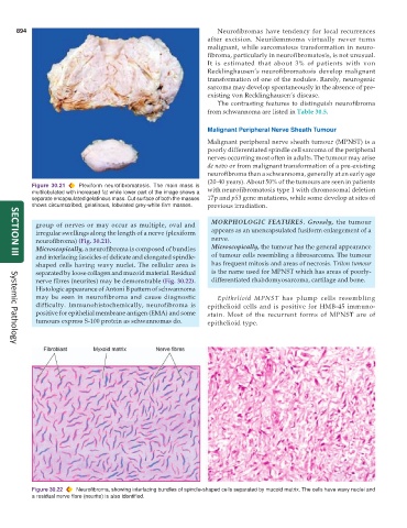
Figure 30.22 Neurofibroma, showing interlacing bundles of spindle-shaped cells separated by mucoid matrix. The cells have wavy nuclei and
a residual nerve fibre (neurite) is also identified.