
Page 907 - Textbook of Pathology, 6th Edition
P. 907
891
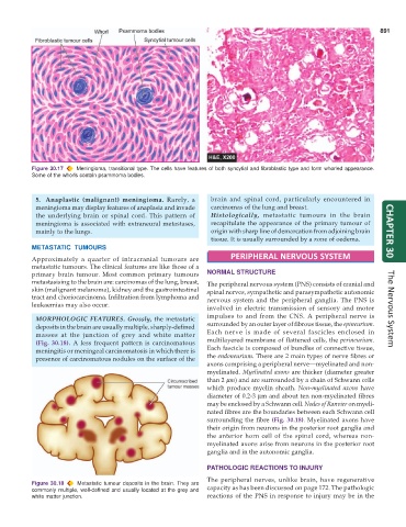
Figure 30.17 Meningioma, transitional type. The cells have features of both syncytial and fibroblastic type and form whorled appearance.
Some of the whorls contain psammoma bodies.
5. Anaplastic (malignant) meningioma. Rarely, a brain and spinal cord, particularly encountered in
meningioma may display features of anaplasia and invade carcinomas of the lung and breast.
the underlying brain or spinal cord. This pattern of Histologically, metastatic tumours in the brain
meningioma is associated with extraneural metastases, recapitulate the appearance of the primary tumour of
mainly to the lungs. origin with sharp line of demarcation from adjoining brain CHAPTER 30
tissue. It is usually surrounded by a zone of oedema.
METASTATIC TUMOURS
Approximately a quarter of intracranial tumours are PERIPHERAL NERVOUS SYSTEM
metastatic tumours. The clinical features are like those of a
primary brain tumour. Most common primary tumours NORMAL STRUCTURE
metastasising to the brain are: carcinomas of the lung, breast, The peripheral nervous system (PNS) consists of cranial and
skin (malignant melanoma), kidney and the gastrointestinal spinal nerves, sympathetic and parasympathetic autonomic
tract and choriocarcinoma. Infiltration from lymphoma and nervous system and the peripheral ganglia. The PNS is
leukaemias may also occur.
involved in electric transmission of sensory and motor The Nervous System
MORPHOLOGIC FEATURES. Grossly, the metastatic impulses to and from the CNS. A peripheral nerve is
deposits in the brain are usually multiple, sharply-defined surrounded by an outer layer of fibrous tissue, the epineurium.
masses at the junction of grey and white matter Each nerve is made of several fascicles enclosed in
(Fig. 30.18). A less frequent pattern is carcinomatous multilayered membrane of flattened cells, the perineurium.
meningitis or meningeal carcinomatosis in which there is Each fascicle is composed of bundles of connective tissue,
presence of carcinomatous nodules on the surface of the the endoneurium. There are 2 main types of nerve fibres or
axons comprising a peripheral nerve—myelinated and non-
myelinated. Myelinated axons are thicker (diameter greater
than 2 μm) and are surrounded by a chain of Schwann cells
which produce myelin sheath. Non-myelinated axons have
diameter of 0.2-3 μm and about ten non-myelinated fibres
may be enclosed by a Schwann cell. Nodes of Ranvier on myeli-
nated fibres are the boundaries between each Schwann cell
surrounding the fibre (Fig. 30.18). Myelinated axons have
their origin from neurons in the posterior root ganglia and
the anterior horn cell of the spinal cord, whereas non-
myelinated axons arise from neurons in the posterior root
ganglia and in the autonomic ganglia.
PATHOLOGIC REACTIONS TO INJURY
The peripheral nerves, unlike brain, have regenerative
Figure 30.18 Metastatic tumour deposits in the brain. They are capacity as has been discussed on page 172. The pathologic
commonly multiple, well-defined and usually located at the grey and
white matter junction. reactions of the PNS in response to injury may be in the