
Page 906 - Textbook of Pathology, 6th Edition
P. 906
890
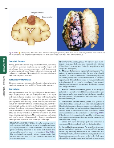
Figure 30.16 Meningioma, The tumour mass is circumscribed (arrow) with irregular surface convolutions and prominent blood vessels. It is
firm in consistency with peripherally adherent thick firm dural tissue. Cut surface of the mass is firm and fibrous.
Germ Cell Tumours Microscopically, meningiomas are divided into 5 sub-
Rarely, germ cell tumours may occur in the brain, especially types: meningotheliomatous (syncytial), fibrous
in children. Common locations are suprasellar region and (fibroblastic), transitional (mixed), angioblastic and
pineal area. Some common examples of such tumours are anaplastic (malignant).
germinoma (seminoma/dysgerminoma), teratoma and 1. Meningotheliomatous (syncytial) meningioma. This
SECTION III
embryonal carcinoma. Morphologically, they are similar to pattern of meningioma resembles the normal arachnoid
their counterparts elsewhere. cap cells. The tumour consists of solid masses of polygonal
cells with poorly-defined cell membranes (i.e. syncytial
TUMOURS OF MENINGES appearance). The cells have round to oval, central nuclei
with abundant, finely granular cytoplasm. Some amount
The most common tumour arising from the pia-arachnoid is
meningioma accounting for 20% of intracranial tumours. of collagenous stroma is present that divides the tumour
into irregular lobules.
Meningioma 2. Fibrous (fibroblastic) meningioma. A less frequent
Meningiomas arise from the cap cell layer of the arachnoid. pattern is of a spindle-shaped fibroblastic tumour in which
Their most common sites are in the front half of the head the tumour cells form parallel or interlacing bundles.
and include: lateral cerebral convexities, midline along the Whorled pattern and psammoma bodies are less common
Systemic Pathology
falx cerebri adjacent to the major venous sinuses features of this type.
parasagittally, and olfactory groove. Less frequent sites are: 3. Transitional (mixed) meningioma. This pattern is
within the cerebral ventricles, foramen magnum, cerebello- characterised by a combination of cells with syncytial and
pontine angle and the spinal cord. Meningiomas are generally fibroblastic features with conspicuous whorled pattern of
solitary. They have an increased frequency in patients with tumour cells, often around central capillary-sized blood
neurofibromatosis 2 and are often multiple in these cases. vessels. Some of the whorls contain psammoma bodies
They are usually found in 2nd to 6th decades of life, with due to calcification of the central core of whorls (Fig. 30.17).
slight female preponderance. Most meningiomas are benign Other forms of degenerative changes like xanthomatous
and can be removed successfully. Rarely, a malignant and myxomatous degeneration may also be encountered,
meningioma may metastasise, mainly to the lungs. in transitional variety.
These first three histologic patterns constitute a
MORPHOLOGIC FEATURES. Grossly, meningioma is spectrum of lesions rather than three distinct entities.
well-circumscribed, solid, spherical or hemispherical mass
of varying size (1-10 cm in diameter). The tumour is 4. Angioblastic meningioma. An angioblastic menin-
generally firmly attached to the dura and indents the gioma includes 2 patterns: haemangioblastic pattern
surface of the brain but rarely ever invades it (Fig. 30.16). resembling haemangioblastoma of the cerebellum, and
The overlying bone usually shows hyperostosis. Cut haemangiopericytic pattern which is indistinguishable from
surface of the tumour is firm and fibrous, sometimes with haemangiopericytoma elsewhere in the body. Both types
foci of calcification. of angioblastic meningiomas have high rate of recurrences.