Page 13 - PRE-U STPM BIOLOGY TERM 1
P. 13

Biology Term 1  STPM  Chapter 2 Structure of Cells and Organelles

                     Exam Tips          Electron microscope
               Remember the basic         1.  Electron microscope is a microscope that uses beams of electrons to
               principles of phase-contrast
               microscope, transmission     form an image.
               and scanning electron
               microscopes including      2.  They are  divided into  transmission electron microscope and
               examples of their uses.      scanning electron microscope.
          2                                 (a)  Transmission electron microscope (TEM)
                                                (i)  It is a microscope that makes use of beams of electrons to
                                                    pass through a very thin section of an object to form an
                                                    image on a monitor screen.
                                2014
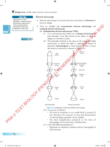
                                                (ii)  The principle involved is the same as the compound light
                                                    microscope in  which the  light is  replaced  by beams of
                                                    electrons;  electromagnet is used instead of lens to focus
                                                    the beams of electrons as shown in Figure 2.6.

                                                                                       Electron
                                                             Light                      source
                                                             source


                                                                                     Electromagnetic
                                                           Condenser
                                                             lenses                    condenser
                                                                                         lens
                                                            Specimen                   Specimen

                                                                                       Objective
                                                            Objective
                                                             lens                        lens


                                                             Ocular                     Ocular
                                                             lens                        lens


                                                             Image
                                                                                        Image

                                                  Light microscope         Electron microscope
                                                    Figure 2.6 Comparison of light and electron microscopes
                                                (iii)  Its uses are as follows:
                                                    •  The limit of resolution is very small which is around 0.5
                                                       nm. Therefore, the structure of virus and ultrastructure
                                                       of cell including organelles can be studied.
                                                    •  The  final  magnification  produced  in  the  form  of
                                                       micrograph can be more than 250,000 times.
                                                (iv)  It has the following limitations:
                                                    •  Experience is required to produce electro-micrograph of
                                                       cell structures.

               58


         02[STPM Bio T1].indd   58                                                                      3/29/18   5:08 PM
   8   9   10   11   12   13   14   15   16   17   18