
Page 46 - PRE-U STPM BIOLOGY TERM 1
P. 46
Biology Term 1 STPM Chapter 2 Structure of Cells and Organelles
(iii) They have thin primary wall. Summary
(iv) They are usually elongated prisms.
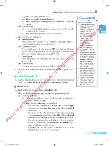
(v) They are living cells with protoplasts and nuclei. They store Specialised plant cells
starch. 1. Parenchyma (thin wall,
(d) Sclerenchyma living with intercellular
(i) It is found in dicotyledonous stems, either on the outside spaces) found in
epidermis, mesophyll,
or inside of the phloem. cortices, pith and 2
(ii) The structure is the same as an ordinary one. ground tissue – for
packing, storage of food
4. The functions are as follows: and photosynthesis
(a) Sieve elements 2. Collenchyma (non-
uniformly thickened
They translocate organic food substances especially sucrose, wall, living and
amino acids, organic acids and proteins. compact) found
(b) Companion cells beneath dicot stem
and leaf epidermis –
They provide energy in the form of ATP and their membranes for strengthening and
have proton pumping-system for the loading of sucrose into the support
3. Sclerenchyma (lignified
sieve tubes from neighbouring mesophyll cells. thick wall fibres or stone
(c) Parenchyma cells) found in phloem
They differentiate to form phloem cells and some remain as of dicot stem, beneath
epidermis in bundle
storage cells. sheath of monocot
(d) Sclerenchyma and in husk or stone of
They protect and support the thin-walled phloem cells. seeds – for protection
and support
4. Xylem (mainly vessel
5. They are found in stems, roots and leaves as shown in Figures 2.26 – element and tracheid of
2.33. thick lignified wall) – for
transport of water and
soluble mineral ions
Specialised Animal Cells 5. Phloem (mainly sieve
tubes and companion
1. Animal cells are classified into four types, based on four fundamental cells of thin wall) – for
tissues i.e. epithelial, nervous, muscle and connective tissues. transport of sucrose
and amino acids.
Epithelial tissues
1. Epithelial tissues are covering or glandular cells.
2. They are divided into covering epithelia and glandular epithelia.
(a) Covering epithelia
(i) Covering epithelia are layers of cells that line the external or
internal surfaces of organs.
(ii) Their structural features are as follows:
• The cells are arranged in a single layer called simple
epithelium or in more than one layer called stratified
epithelium.
• The shape of the cells depends on the types; scale-like
called squamous epithelium, cube-like called cuboidal
epithelium and column-like called columnar epithelium.
• The cells are attached to a thin layer of fine connective
tissues at the bottom called basement membrane. This
helps to attach it to other tissues.
• At the top, the cells are exposed to air such as in the skin,
or liquid such as those of the stomach.
91
02[STPM Bio T1].indd 91 3/29/18 5:08 PM